Developmental Biology Program
The Mary Baylies Lab
Research

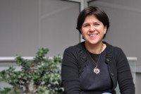
Mary Baylies, PhD
Developmental biologist Mary Baylies studies the mechanisms that form and maintain muscle both during normal development and in disease.
Research Projects

Featured News
In the Lab
A new study in flies reveals a previously unknown type of cooperation at work in muscle cells.
Learn about Mary Baylies, a developmental biologist studying muscle biology at the Sloan Kettering Institute.
More than 500 high school students and their teachers filled the Rockefeller Research Laboratories to learn about recent discoveries.
People
Mary Baylies, PhD
- The Baylies laboratory studies the mechanisms that form and maintain muscle both during normal development and in disease.
- PhD, The Rockefeller University
- [email protected]
- Email Address
- 212-639-5888
- Office Phone
- Download CV
- PDF File
Members


Member
Research Technician

Graduate Student

MSK Engage scholar

Graduate Student

Administrative Assistant
Postdoctoral Research Scholar
Graduate Student

WCGS Graduate Student

GSK Graduate Student

Senior Research Scientist

Postdoctoral Research Scholar

Postdoctoral Research Scholar


Research Fellow
Graduate Student

Research Fellow
Research Fellow
Graduate Fellow
Research Fellow
Graduate Fellow
Graduate Student

Graduate Research Assistant

Graduate Student

Research Fellow
Graduate Student

Graduate Student
Research Technician

Postdoctoral Research Fellow

Research Fellow
Graduate Student
Research Technician

MSK Bridge Program Scholar

Graduate Student
Graduate Student
Achievements
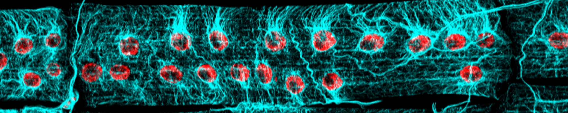
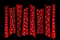
- Identified that global, regional, and local inputs contribute to nuclear size regulation in multinucleated skeletal muscle fibers.
- Demonstrated that properly placed nuclei are critical for healthy muscle function.
- Discovered that microtubules are critical for the movement and positioning of nuclei in the multinucleated skeletal muscle cell.
- Developed Drosophila models of muscle wasting in cancer cachexia and aging.
- Identified how actin filament dysregulation contributes to skeletal muscle dysfunction in models of nemaline myopathy.
- Developed a Drosophila model for metastasis in alveolar rhabdomyosarcoma, a type of soft tissue cancer most often seen in children.
- Described how nuclear polarization occurs during myonuclear movement in vivo.
- Completed the first spatial and temporal characterization of an F-actin structure that forms at the myoblast fusion site.
- Identified transcription factors and chromatin regulators that are critical for muscle identity.
- Identified 3-D arrangement of myoblasts that occur during muscle specification and morphogenesis.
- Discovered signal transduction pathways (Notch, Wnt, BMP, and RTK) and transcription factors that are critical for muscle identity.
- President of the Society for Muscle Development (2009-2013).
- Frederick Adler Chair for Junior Faculty (1997-2003).


