Molecular Biology Program
The Prasad Jallepalli Lab
Research

Prasad Jallepalli, MD, PhD
Member
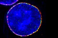
Our laboratory is interested in how human cells accurately duplicate and transmit their chromosomes during each round of division.

Featured News
Snapshot
The discovery of a molecular process that slows down cell division could provide new understanding about how some cancers develop.
People
Prasad Jallepalli, MD, PhD
Member
- Molecular biologist Prasad Jallepalli studies the mechanisms that ensure accurate chromosome transmission in human cells.
- MD, PhD, Johns Hopkins University School of Medicine
- [email protected]
- Email Address
- 212-639-3252
- Office Phone
Members


Member & Professor
Senior Research Scientist
Research Technician


Research Scholar

Graduate Student

Research Scholar

Research Assistant

Research Associate
Graduate Student
Graduate Student
Research Fellow
Graduate Student
Research Fellow
Research Fellow
Research Fellow

Graduate Student
Vice President, Bipharma Equity Research, Wells Fargo

Research Associate
Graduate Student
Research Fellow
Achievements
- Phi Beta Kappa, Harvard University (1992)
- M.D./Ph.D. Fellow, Medical Scientist Training Program, Johns Hopkins University (1992-1999)
- Postdoctoral Fellow, Damon Runyon Cancer Foundation (1999-2002)
- Scholar, V Foundation for Cancer Research (2003)
- Frederick R. Adler Chair for Junior Faculty (2003-2007)
- Pew Scholar in the Biomedical Sciences (2003-2007)
- Louise & Allston Boyer Young Investigator Award (2008)
- Research Scholar, American Cancer Society (2008-2011)