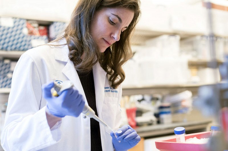
Neurologist Dr. Adrienne Boire in her lab
Unique markers of inflammation and neurodegeneration in cerebrospinal fluid were present several weeks after initial SARS-CoV-2 infection in cancer patients with neurologic symptoms. These inflammatory cytokines were absent in plasma and likely drove the neurologic dysfunction, according to our research published recently in Cancer Cell. (1)
Acute SARS-CoV-2 infection is frequently associated with neurologic symptoms, such as headache, early anosmia, and dysgeusia, that usually resolve. (2) However, patients with more severe illness may experience protracted delirium, seizures, and meningoencephalitis. (3) Understanding the pathophysiology of the neurologic consequences of COVID-19 in cancer patients is crucial because these patients have a higher risk of severe illness with COVID-19 due to their immunocompromised state and low functional reserve. (4)
We prospectively evaluated a series of cancer patients with moderate to severe neurologic sequelae after SARS-CoV-2 infection, including a targeted proteomics analysis of cerebrospinal fluid (CSF). We found leptomeningeal inflammatory cytokines — in the absence of viral neuroinvasion — nearly two months after the onset of respiratory symptoms. Most of these inflammatory mediators were driven by type II interferon, known to cause neuronal injury in other diseases. We also discovered elevated intracranial levels of matrix metalloproteinase-10 (MMP-10) that correlated with the degree of clinical neurologic dysfunction. (1)
Our findings suggest that early neurologic assessments are essential, and anti-inflammatory treatments, such as dexamethasone, may be beneficial for managing neurologic complications in cancer patients with severe or prolonged COVID-19. Further, MMP-10 deserves further study as a prognostic biomarker of neurologic dysfunction. (1)
At MSK, we are dedicated to improving patients’ outcomes through leading-edge research and cancer care. Our researchers and clinician-scientists continue to play an important role in the international response to the COVID-19 pandemic.
How Does COVID-19 Affect the CNS?
Large studies suggest that the neurologic complications of COVID-19 are far more prevalent than originally thought, occurring in 7 to 69 percent of patients with severe infection. (5) However, the mechanism by which COVID-19 affects the central nervous system (CNS) is unclear. Hypotheses include direct viral invasion, toxicity from systemic cytokine release syndrome (CRS), or a combination of both.
Studies to date regarding the potential for SARS-CoV-2 to invade the CNS lack consensus. Detectable virus in CSF has only been found in a small number of patients with neurologic symptoms. (3), (6), (7), (8) Small case reports of up to three patients have reported elevated levels of proinflammatory cytokines in the spinal fluid of acutely infected patients, including interleukin (IL)-6, C-X-C motif chemokine ligand (CXCL)-10 (also called interferon-induced protein-10), and C-C motif chemokine ligand (CCL)-2. (7), (9), (10)
No studies to date have characterized the full extent and duration of neuroinflammatory response to COVID-19 in a large patient population, nor compared the degree of neuroinflammation with the severity of neurologic dysfunction.
Study Design
Between May and July 2020, we prospectively evaluated 18 cancer patients with confirmed SARS-CoV-2 respiratory infection who subsequently developed moderate to severe neurological symptoms.
We also examined whether CSF composition correlated with the degree of neurologic dysfunction.
We compared CSF from COVID-19-positive cancer patients with three cancer patient cohorts from the pre-pandemic era to control for other cancer-related causes of neuroinflammation: patients with neurotoxicity associated with brain metastases; patients who experienced immune effector cell-associated neurotoxicity syndrome (ICANS) (11) after chimeric antigen T cell (CAR T) cell therapy; and patients with autoimmune encephalitis. (12), (13)
Study Results
Clinical characterization of COVID-19-related neurologic sequelae in cancer patients
Primary cancers among our COVID-19-positive cohort included a wide range of solid tumors and hematologic malignancies. Thirteen patients (72 percent) received tumor-directed treatment within 30 days of COVID-19 onset, and seven (39 percent) were immunocompromised before infection. Medical comorbidities were common: hypertension (56 percent), smoking history (44 percent), hyperlipidemia (33 percent), diabetes (28 percent), and previous ischemic infarct (six percent). (1)
Sixteen patients had SARS-CoV-2 infection confirmed by nasopharyngeal swab. We included two additional patients based on positive serum SARS-CoV-2 antibody screen and recent illness consistent with COVID-19. All 18 patients presented with classic COVID-19 symptoms, including dyspnea and cough. Many had elevated ferritin, D-dimer, IL-6, and C-reactive protein. Eleven patients experienced severe hypoxic respiratory failure and systemic inflammatory response syndrome requiring mechanical ventilation. (1)
Patients exhibited a wide range of neurological symptoms after SARS-CoV-2 infection, with all showing at least a mild dysexecutive syndrome consistent with global encephalopathy. Additional neurological diagnoses included prolonged critical care delirium (10 patients), limbic encephalitis (four patients), refractory headaches (two patients), rhombencephalitis (one patient), and large territory infarctions (one patient). Sedation holidays confirmed that persistent neurologic impairment was out of proportion to critical care delirium. The median delay between the onset of respiratory symptoms and clinically documented neurologic sequelae was 19 days (range 0 to 77 days). (1)
Neurologic testing included bedside examination as well as magnetic resonance imaging or head computed tomography. Three patients showed encephalitic changes in the form of non-enhancing T2-hyperintense white matter and cortical changes affecting the cerebellar or limbic structures. We also noted instances of large territory infarcts, diffuse microhemorrhages, and increased subcortical or periventricular white matter disease compared to pre-COVID-19 imaging. Diffuse bihemispheric slowing was the most common finding in 12 patients and was irrespective of receiving sedation. (1)
CSF composition after COVID-19
Thirteen patients underwent spinal fluid collection at a median of 57 days (range 5 to 142) from the onset of respiratory symptoms and 37 days (range 1 to 117) from the onset of neurologic symptoms. Spinal fluid collection was conducted when neurologic symptoms could not be attributed to other non-infectious causes, and the benefits of CSF testing outweighed the procedural risks. (1)
Interestingly, cell count, protein, and glucose levels were normal. Two patients with known CNS metastases had measurable pleocytosis. CSF immune cell differentials were lymphocyte predominant in 77 percent and monocyte predominant in 23 percent of patients. We detected oligoclonal bands in both the serum and CSF of 83 percent of patients, indicating systemic rather than intracerebral production of gamma globulins. Only two patients had elevated intracranial pressure and abnormal protein levels, which were most likely due to active brain metastases rather than neurologic injury from COVID-19. (1)
We optimized a polymerase chain reaction (PCR)-based assay to test CSF and used a commercially available ELISA to characterize SARS-CoV-2 neuroinvasion and test for the presence of viral RNA and CSF structural proteins. No patients had detectable levels of virus or N or S structural proteins in their CSF. (1)
Proteomic analysis of neuroinflammation
Given that direct viral invasion did not explain the prolonged neurologic dysfunction, we tested CSF from ten patients using an extensive proteomics assay to look for patterns of inflammation and neuronal damage.
We identified a significant accumulation of 12 inflammatory mediators in the CSF of COVID-19 patients, with levels approaching those seen in patients with severe ICANS. These mediators were: IL-6 and -8, interferon (IFN)-γ, CXCL-1, -6, -9, -10 and -11, CCL-8 and -20, MMP-10, and eukaryotic translation initiation factor 4E binding protein 1 (4E-BP1). All of these mediators are associated with cytokine and chemokine signaling, immune cell function, senescence, and neuroinflammation.
Using an inflammatory signature of these 12 mediators, we found a distinct increase in inflammatory signalling in COVID-19-positive patients compared to all COVID-10-negative controls (p = 0.029). An analysis of matched plasma and CSF from 12 COVID-19 patients revealed that IFN-ß and IL-8 appeared to be produced predominantly within the leptomeninges, whereas levels of IFN-α, -γ, and -λ, and chemokines CXCL-9 and CXCL-11 were enriched in plasma. (1)
Comparing levels of these 12 inflammatory markers in CSF with clinical scores of neurologic dysfunction at the time of spinal tap revealed that CSF levels of MMP-10 correlated with the degree of neurologic insult (1) as measured by the Karnofsky performance status and the disability rating scale. (14)
Diagnostic and Treatment Implications
The specific mechanisms involved in COVID-19 inducing neuroinflammation and the extent of neuronal damage in cancer patients merit further investigation. The correlation between MMP-10 and neurologic dysfunction is a prognostic biomarker that deserves further study in cancer patients experiencing prolonged neurologic issues after COVID-19. (1)
In the meantime, our results have important diagnostic and treatment implications. The proinflammatory CSF cytokines we identified were elevated in the weeks following respiratory illness. These cytokines can cause neuronal damage similar to other encephalitides, suggesting that early assessments are crucial.
Dexamethasone, a potent steroid and immunosuppressant, is the only intervention that has demonstrated reductions in deaths in hospitalized patients with severe COVID-19. (15) The impact of this drug on COVID-19-related neurologic toxicity has not yet been defined. However, we found that the neurologic toxicity in COVID-19 patients was biochemically similar to CAR T cell neurotoxicity, which is currently managed through early diagnosis and use of anti-inflammatory agents, including dexamethasone. (16), (17), (18)
Advancing Cancer Care Through Translational Research
We are deeply grateful to the patients and families who donated clinical samples and contributed to our research during the COVID-19 pandemic.
My lab within the Human Oncology & Pathogenesis Program at MSK focuses on leptomeningeal and parenchymal metastases to the central nervous system (CNS). We employ multiple complementary approaches to identify and target cancer cell adaptations to the challenging microenvironmental constraints posed by the CNS. Our goal is to use cutting-edge translational research to advance the development of new treatment options for cancer patients.
The study was supported by the National Cancer Institute/MSK Support Grant P30 CA008748); and the Pew Charitable Trusts GC241069, the Damon Runyon Cancer Research Foundation GC240764, and the Pershing Square Sohn Cancer Research Alliance GC239280 (Dr. Boire.). First author and research fellow Jan Remsik, PharmD, PhD, is supported by the American Brain Tumor Association Basic Research Fellowship, the Terri Brodeur Breast Cancer Foundation Fellowship, and the Druckenmiller Center for Lung Cancer Research.
Dr. Boire is an inventor on the United States Provisional Patent Application no. 62/258,044 “Modulating Permeability of The Blood Cerebrospinal Fluid Barrier, “an unpaid member of the Scientific Advisory Board of EVREN Technologies, and a paid consultant for Kite Gilead, Juno/Celgene, Janssen, and In8bio. All other authors declared no competing interests.

