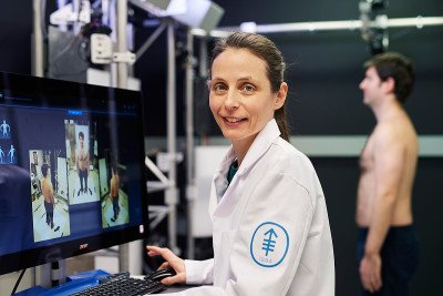
Daphne Demas is a senior medical photographer whose work helps screen patients for melanoma — and spare them unnecessary skin biopsies.
What is My Risk for Melanoma?
A risk factor is anything that increases your chance of getting a disease, such as cancer.
Learn about the risk factors for melanoma.
Melanoma Screening for People With Symptoms or Risk Factors
MSK melanoma experts recommend regular screening for people who have symptoms, or are at higher risk for melanoma. This includes people who have had treatment for melanoma. Regular screening will help find melanoma early, when it’s easier to treat. Melanoma is a serious condition and finding it early is very important.
Learn about the symptoms of melanoma.
We recommend you make regular appointments with your dermatologist (skin doctor) to check for melanoma if you have:
- A family history of melanoma in 2 or more relatives related to you by blood.
- Many moles or atypical (dysplastic) moles. Moles are benign (not cancer). Atypical moles can have uneven borders or be different colors. They also can be asymmetrical (they don’t look the same on all sides).
- Many actinic keratoses spots. These are grey or pink scaly patches of skin in areas often exposed to the sun that may become cancer (precancerous lesions).
- A personal history of many basal cell or squamous cell skin cancers.
What Happens During a Skin Cancer Screening?
Your healthcare provider will look for new growths, spots, or bumps on your skin. They will look for anything on your skin that may be cancerous or precancerous.
If they find an area that does not look normal, they may recommend more tests. They may do a skin biopsy to check for cancer. A biopsy is a procedure done to take samples of tissue to check for cancer.
Melanoma Screening and Surveillance at MSK
People at a higher risk for melanoma can get checked for the disease by an MSK dermatologist.
We use several imaging methods to look for skin cancer, including:
- 3D whole body imaging, to monitor changes in your skin over time.
- Dermoscopy, which uses a hand-held instrument to see below the top layer of skin.
- Confocal microscopy, a new method to diagnose cancer that looks at structures in your skin at a cellular level.
3D whole body imaging at MSK
MSK is a leader in 3D whole body imaging. This imaging system uses more than 90 cameras to take pictures of the whole body. We use the images to make a digital 3D model on a computer screen.
This model shows every mole or spot on your body. Doctors can zoom in to get a closer look. The scan takes much less time than traditional photography. It only takes a few minutes for a full session, compared with more than an hour for the older 2D version.
These 3D tests may help your doctor spot melanoma early, when it’s easier to remove and cure. They are very helpful for people who have many moles or atypical moles. Our screening tools also can help you avoid a skin biopsy that is invasive. An invasive procedure means we have to cut the body.
MSK offers melanoma surveillance services, including 3D whole body imaging for high-risk people, at our dermatology locations:
Request an Appointment
Available Monday through Friday, to (Eastern time)

