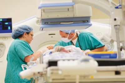
Memorial Sloan Kettering's Center for Image Guided Intervention has two devices for performing CT angiography to visualize and plan treatments.
Image-guided interventions are revolutionizing cancer diagnosis and treatment. MSKCC’s new Center for Image-Guided Intervention (CIGI), which opened in June, offers cancer patients the most advanced, minimally invasive diagnostic and treatment options in a unique multidisciplinary setting designed to foster rapid innovations in cancer care.
Image-guided interventions allow physicians to see and perform procedures inside the body without making large incisions. Interventional radiologists utilize sophisticated imaging methods to diagnose cancers and treat them in minimally invasive ways, such as applying heat or cold, blocking blood vessels that feed tumors, or delivering chemotherapy and radiation directly to tumors. Image-guided approaches to cancer care can reduce risks, shorten hospital stays, and decrease the cost to patients.
The recently opened facility at MSKCC includes not only CIGI but an expansion of the Surgical Day Hospital and a new endoscopy suite. The close proximity of these three entities will allow interventional radiologists, surgeons, and endoscopists to collaborate closely in developing new procedures and treatments. The new 40,000-square-foot facility cost more than $100 million in construction and equipment. Approximately 120 patients a day are expected to be seen in the facility.
“Seven years in the making, this magnificent facility is a result of shared vision and collaboration,” said Hedvig Hricak, Chair of the Department of Radiology, who, along with Peter T. Scardino, Chair of the Department of Surgery, was instrumental in conceptualizing and planning the new facility. “The close proximity of radiology, surgery, and endoscopy will produce innovations that advance the medicine of the 21st century and also provide a superb environment for our patients.”
The tools of interventional radiology (IR) physicians include fluoroscopy, computed tomography (CT), ultrasound, positron emission tomography (PET), and magnetic resonance imaging (MRI). CIGI has six procedure rooms that contain leading-edge imaging equipment.
MSKCC has one of the first combined “angio-CT” suites in the United States, and the equipment has proved so valuable for procedures and treatments that CIGI contains two additional angio-CT units. These combination rooms have enabled procedures that have never been performed before. Traditionally, interventional procedures were done using conventional two-dimensional x-ray equipment. However, three-dimensional CT, MRI, and PET display anatomy in much greater detail and provide additional information about metabolism and physiology that improves cancer detection and characterization. All three of these 3D diagnostic imaging techniques are available for guiding interventional procedures in the new facility.
“CIGI is an unbelievable platform for imaging and treating cancer,” said Stephen B. Solomon, Interventional Radiology Service Chief and Co-Chair of CIGI. “We have an interventional PET/CT scanner in the center that will allow us to take advantage of new markers and tracers to pinpoint cancers in ways that were impossible before. The two new MRI rooms will enable us to continue refining a new approach in which we use MRI not as a diagnostic tool but to guide and monitor therapies in real time. For example, we are developing ways to noninvasively destroy tumors with heat by using focused ultrasound technology, and minimally invasively by using lasers, and the MRIs enable us to closely monitor the temperature to ensure we are destroying the target.”
Dr. Scardino explained that the new facility also makes it possible to consolidate several procedures performed by different specialists into a single patient visit. “A patient with an unspecified mass in the chest could typically require a CT scan by an interventional radiologist to biopsy it, an ultrasound by an endoscopist to stage it and determine its size, and a surgical procedure called a mediastinoscopy to take out a lymph node,” he said. “In the past, getting all three procedures would require three separate trips to the hospital over several weeks. Now we can do them consecutively in the same day and move promptly to therapy.”
In addition to having the most modern technology, the new facility offers amenities that enhance the patient experience, including privacy. For example, a patient preparation area consisting of 48 individual private spaces called “bays” contain equipment to monitor vital signs and administer intravenous fluids, as well as a computer so medical staff can complete documentation without leaving the patient’s bedside.
CIGI was designed with the understanding that interventional radiology will continue to progress rapidly, making it difficult to predict how new technology might be implemented in the future. Procedure rooms were configured to have extra space for new devices that may be developed in upcoming years.
“The rooms are designed to provide much more than conventional IR rooms,” Dr. Hricak said. They are furnished with flat-panel displays — called the “Walls of Knowledge” — that include all patient clinical and laboratory data. In addition, a state-of-the-art communication system allows physicians in separate CIGI procedure rooms to consult via videoconferencing and share real-time images.
Along with the clinical facility there is a dedicated CIGI laboratory research facility devoted to developing and testing phantoms and animal models techniques that can eventually be evaluated in clinical trials.
