Neurovascular Uncoupling and Cortical Reorganization in Brain Tumors
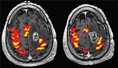
Dr. Holodny was the first to describe the impacts of neurovascular uncoupling in fMRI of brain tumors. The blood-oxygenation level-dependent (BOLD) fMRI signal relies on hemodynamic changes from autoregulation in activating brain areas. However, the presence of a tumor and its abnormal neovasculature causes the brain to lose its ability for autoregulation, leading to false-negative behavior caused by neurovascular uncoupling. Such false-negative behavior may lead to inadvertent resection in eloquent brain areas. Our work hopes to not only characterize this limitation but also discover strategies to improve the accuracy of fMRI.
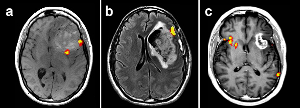
The presence of a tumor can also trigger cortical reorganization, which is when the brain relocates its functional activation from the damaged cortical area to another region. This process is believed to be a means for the brain to compensate for the loss of activation around the tumor. Our work has particularly focused on understanding contralateral brain reorganization, which is the transfer of function to the contralateral hemisphere.
- Holodny AI, Schulder M, Liu WC, Maldjian JA, Kalnin AJ. Decreased BOLD functional MR activation of the motor and sensory cortices adjacent to a glioblastoma multiforme: implications for image-guided neurosurgery. AJNR Am J Neuroradiol. 1999. 20(4):609-12.
- Shaw K, Brennan N, Woo K, Zhang Z, Young R, Peck KK, Holodny AI. Infiltration of the basal ganglia by brain tumors is associated with the development of co-dominant language function on fMRI. Brain Lang. 2016. 155-156:44-8.
- Cho NS, Peck KK, Zhang Z, Holodny AI. Paradoxical activation in the cerebellum during language fMRI in patients with brain tumors: possible explanations based on neurovascular uncoupling and functional reorganization. The Cerebellum. 2018. 17(3):286-293.

BOLD fMRI activation during a bilateral hand motor task is decreased in ipsilateral areas around the tumor compared to the contralateral side. This may be the result of neurovascular uncoupling false-negative behavior caused by the tumor. |

Language tasks are typically left-dominant for right-handed patients (a, b), but right-dominant activation can also occur, likely due to contralateral reorganization (c). |
Advanced MRI Methods: Imaging Techniques and Models of Complex Neurological Systems
We are also exploring the clinical applications of other advanced imaging methods for patients with brain tumor. Some of these methods include diffusion tensor imaging (DTI), dynamic contrast-enhanced (DCE) perfusion MRI, and resting-state fMRI. Our work has elucidated biomarkers for evaluating tumors and their treatment response that are more accurate than those in conventional imaging, which has guided the implementation of these advanced imaging methods into clinical care at our institution.
- Arevalo-Perez J, Peck KK, Young RJ, Holodny AI, Karimi S, Lyo JK. Dynamic contrast-enhanced perfusion MRI and diffusion-weighted imaging in grading of gliomas. J Neuroimaging. 2015. 25(5):792-8.
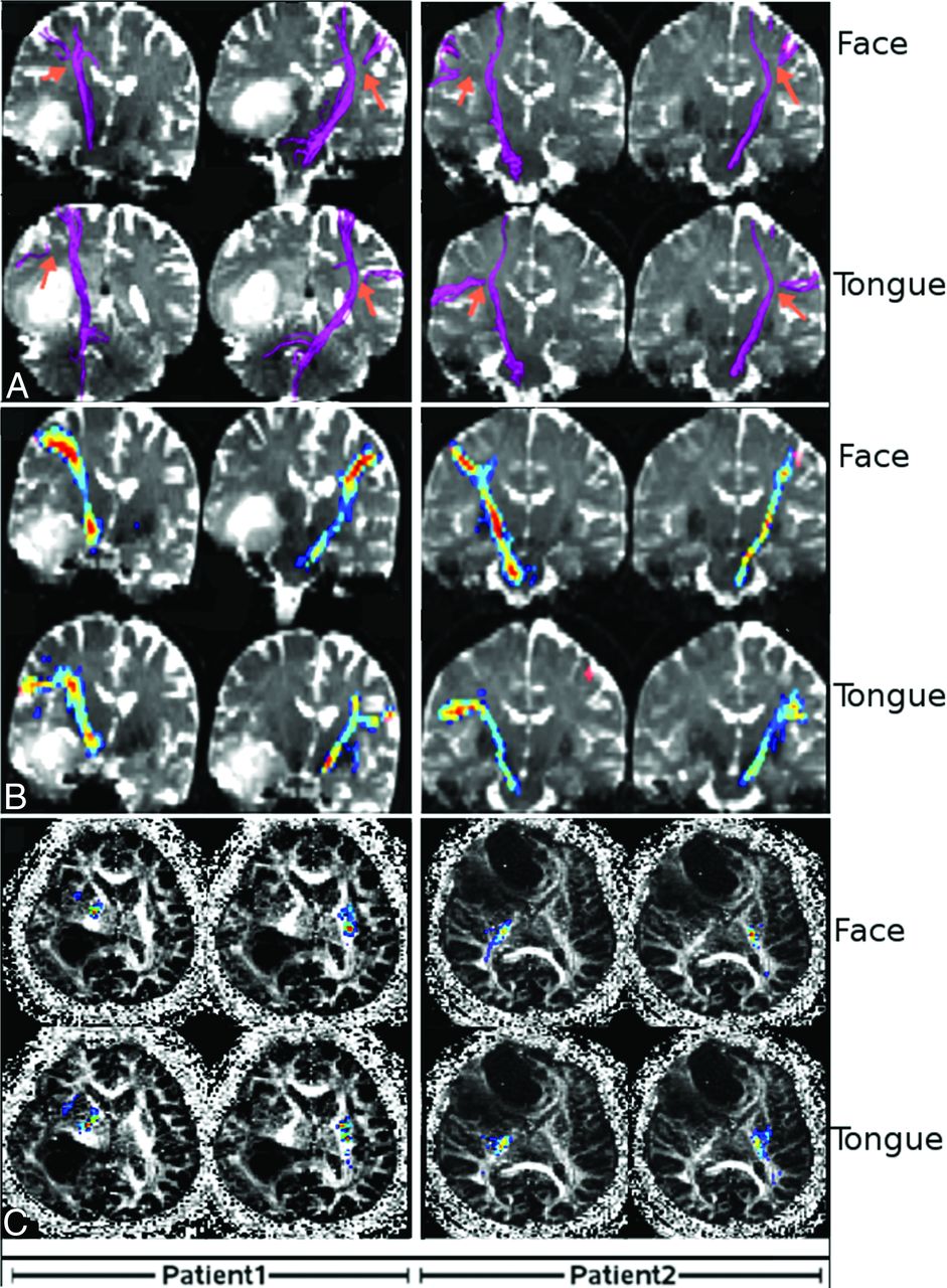
- Jenabi M, Peck KK, Young RJ, Brennan N, Holodny AI. Identification of the corticobulbar tracts of the tongue and face using deterministic and probabilistic DTI fiber tracking in patients with brain tumor. AJNR Am J Neuroradiol. 2015. 36(11):2036-41.
- Mallela AN, Peck KK, Petrovich-Brennan NM, Zhang Z, Lou W, Holodny AI. Altered resting-state functional connectivity in the hand motor network in glioma patients. Brain Connect. 2016. 6(8):587-95.
- Hamidian S, Vachha B, Jenabi M, Karimi S, Young RJ, Holodny AI, Peck KK. Resting-state functional magnetic resonance imaging and probabilistic diffusion tensor imaging demonstrate that the greatest functional and structural connectivity in the hand motor homunculus occurs in the area of the thumb. Brain Connect. 2018. 8(6):371-9.

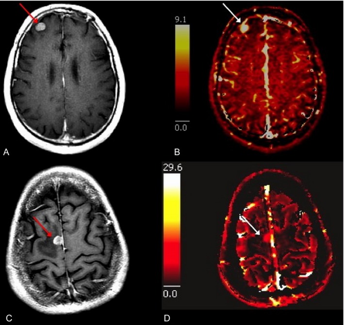
Conventional MRI (left) shows similar appearances for melanoma brain metastasis (top) and lung cancer brain metastasis (bottom) while dynamic contrast-enhanced MRI (right) can differentiate them from plasma volume (Vp) maps.
|

Face and tongue motor tracts generated using FACT (a) and probabilistic (b, c) fiber tractography. |
Grants
NIH-NIBIB 1R01EB022720-01 “Graph theoretical analysis of the effect of brain tumors on functional MRI networks”
The main goal is to develop a software tool that will allow end-users from the broad neuroscience community to identify and analyze the most influential parts of the brain in various disease states. The tools developed by this project will aid in the understanding, diagnosis, and therapy of brain disorders thought to be due to disruptions of brain connectivity (e.g. brain tumors, Alzheimer’s disease, ADHD, stokes or traumatic brain injury).
NIH-NCI 1 R21 CA220144-01 “Identification of Essential Areas of the Brain in Pre-Operative Brain Tumor Patients Using BOLD fMRI and Independent Physiological Parameters.”
The main goal is to overcome the problem of neurovascular uncoupling in fMRI of brain tumors, by incorporation of an independent measurement of cerebrovascular reactivity using breath-holding MRI (BH MRI) to calibrate the BOLD response, which will improve the accuracy of BOLD fMRI in the vicinity of brain tumors and have an immediate, positive impact on brain tumor resections.
NIH-NCI R25CA020449 “Summer Research Experiences for Medical Students Supervised by Faculty Mentors”
The goal is to address a significant challenge in the field of oncology: to build an oncology workforce that is both large enough and diverse enough to meet the needs of the aging population.
Bayer Pharmaceuticals “Evaluation of Metastatic Spinal Bone Marrow Response to Radiation Therapy Using T1 weighted Dynamic Contrast-Enhanced Perfusion”
The goal of this study is to explore if DCE-MRI parameters change from baseline to post treatment scans. This is a pilot study with the eventual goal of additional larger studies to assess if DCE-MRI has prognostic value and can detect treatment success and failure earlier than conventional MRI.
The Dana Foundation “Hyperpolarized Pyruvate MRI to characterize Glioma Metabolism Non-invasively in man.”
This study aims to test the application of a small hyperpolarized metabolite, pyruvate, to brain tumor imaging. This study will be the first systematic application of hyperpolarized MRI to brain tumor imaging.