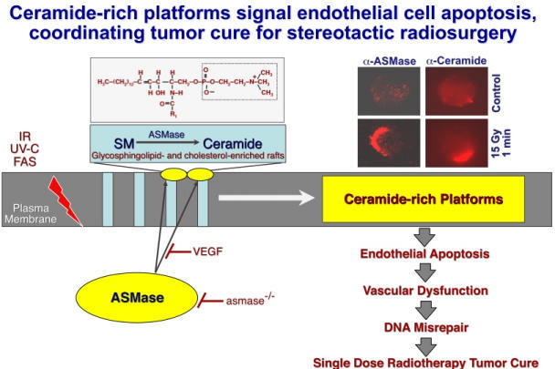
Our laboratory focuses on the role of sphingolipid signaling as a stress response. In this pathway, generation of the second messenger ceramide in response to diverse environmental and pharmacologic stresses (heat, ionizing radiation, ultraviolet light, chemotherapeutic agents, oxidative challenges, etc.) occurs either by degradation of sphingomyelin or by de novo synthesis. The quality and quantity of the ceramide response, in combination with other signals, determines whether adaptation or apoptosis ensues. This pathway is evolutionarily conserved and is obligate for the heat shock response in yeast.

Our program uses a multidisciplinary approach involving genetics, biochemistry, and cell biology to address these issues. Knockouts of relevant genes are generated in mice and concomitantly in the nematode Caenorhabditis elegans. Studies in C. elegans provide information regarding the molecular ordering of ceramide signaling during stress and development, whereas studies in mice provide information of tissue/organ distribution of ceramide effects. The goal of these studies is to determine under what conditions the ceramide signal is involved in normal physiology and in pathophysiology of disease and the development of pharmacologic agents to suppress or enhance signaling through this system in vivo. Evidence suggests that the sphingomyelin pathway is critical for ionizing radiation-induced death of oocytes and endothelium, is co-opted by a variety of microorganisms as a mechanism to gain access to the cellular interior, and serves as an important co-signal for TNF receptor superfamily-induced death. Pre-clinical data suggest that the therapeutic success of stereotactic radiosurgery, a new form of high dose radiotherapy capable of curing radioresistant cancer, depends on ceramide-mediated apoptosis of the tumor microvascular network (see Figure 1).