Early detection of melanoma is paramount to determine the best treatment approach and achieve a potential cure.
Advances in diagnostic and imaging modalities are making it possible to detect melanoma earlier, with greater accuracy, and are helping guide surgical approaches and patient monitoring plans.
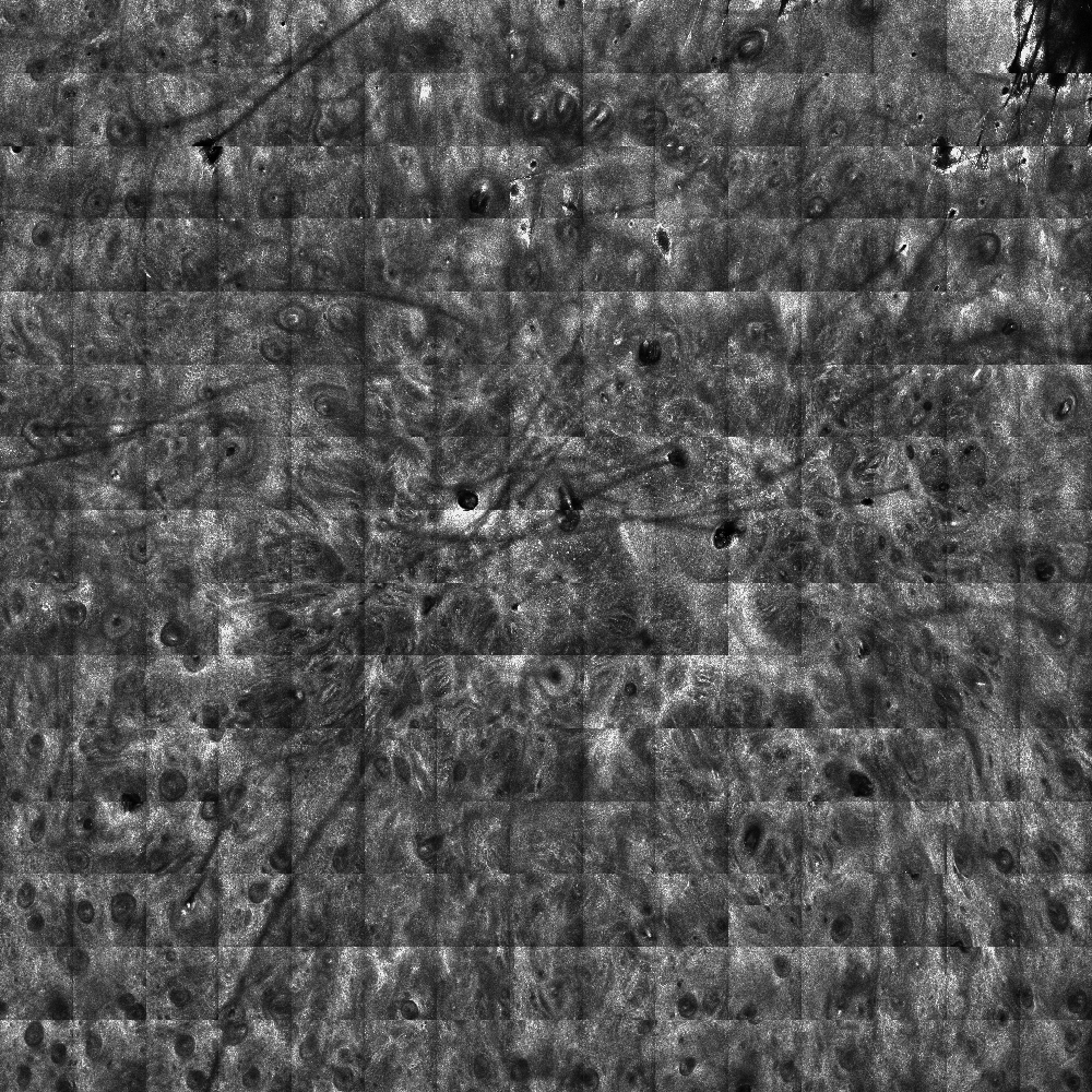
One such recent advance, used by the Dermatology Service at Memorial Sloan Kettering Cancer Center, is reflectance confocal microscopy or RCM.
RCM for Better Detection
RCM is an exciting, non-invasive imaging tool that provides improved accuracy in melanoma detection. The technology uses a low-power laser to provide real-time imaging of the epidermis and superficial papillary dermis. It captures the natural reflectivity of different cellular structures, including melanocytes, and can be quite useful to diagnose benign and malignant lesions. (1),(2),(3),(4)
Since it is noninvasive, RCM allows us to reimage the same area over time without altering the tissue.
Complex Melanoma Challenges
Multiple forms of cutaneous melanoma can pose diagnostic and therapeutic challenges. For example, lentigo maligna (LM) is an early form of melanoma in situ. Diagnosis can often be delayed due to poorly defined and irregular borders, subclinical extension, and its location on a background of severely sun-damaged skin. If delayed in diagnosis it can continue to develop into invasive lentigo maligna melanoma.
For LM occurring on the head and neck, or in other anatomically sensitive areas, it can be difficult to decipher where the lesion boundaries extend, making it difficult to determine how much tissue to resect. Further, if we treat this in situ disease with non-surgical therapies, it can be difficult to assess success or failure.
One of the disadvantages of RCM is a small field of view and limited depth. However, in our recently published study, we tested the accuracy and specificity of RCM to diagnose challenging melanocytic lesions. We studied 17 patients with known or suspected LM by evaluating a total of 63 skin sites with RCM. The sites were then biopsied to compare the RCM diagnosis with diagnosis by histopathology. When LM was present, as confirmed by biopsy, RCM detected it 100 percent of the time. When LM was absent, RCM found it was absent 71 percent of the time. (5)
Improved Surgical Approaches
We have also used RCM to assist in surgical resection of large, complex facial melanoma. We have found that it helps to delineate subclinical extension before surgery, allowing us to achieve clear margins in one operation. (6)
We will continue to use the latest imaging technologies, including RCM in conjunction with other imaging techniques, to better diagnose melanoma lesions earlier and to help plan treatment approaches for patients with complex head and neck lesions.
- Guitera P, et al. The impact of in vivo reflectance confocal microscopy on the diagnostic accuracy of lentigo maligna and equivocal pigmented and nonpigmented macules of the face. J Invest Dermatol 2010; 130(8):2080-91.
- Guitera P, et al. In vivo reflectance confocal microscopy enhances secondary evaluation of melanocytic lesions. J Invest Dermatol 2009; 129(1):131-8.
- Langley RG, et al. The diagnostic accuracy of in vivo confocal scanning laser microscopy compared to dermoscopy of benign and malignant melanocytic lesions: a prospective study. Dermatology 2007; 215(4):365-72.
- Pellacani G, et al. The impact of in vivo reflectance confocal microscopy for the diagnostic accuracy of melanoma and equivocal melanocytic lesions. J Invest Dermatol 2007; 127(12):2759-65.
- Menge T, Hibler B, Cordova M, Nehal K, Rossi AM. Concordance of handheld reflectance confocal microscopy (RCM) with histopathology in the diagnosis of lentigo maligna (LM): A prospective study. J Am Acad Dermatol 2016; 74(6):1114-20.
- Hibler BP, Cordova M, Wong RJ, Rossi AM. Intraoperative real-time reflectance confocal microscopy for guiding surgical margins of lentigo maligna melanoma. Dermatol Surg 2015; 41(8):980-3.
