An innovative 3D motion management platform for guiding stereotactic body radiation therapy (SBRT) significantly reduced late urinary toxicity in patients treated for prostate cancer compared to a commercial image-guidance system, according to study results presented October 2 at the ASTRO 2023 Annual Meeting in San Diego. (1)
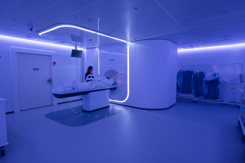
MSK medical physicists and radiation oncologists developed the 3D megavoltage (MV) and kilovoltage (kV) image guidance (MKIG) platform to replace a commercial approach that used 2D intrafraction motion review (IMR). MSK began using the new MKIG in 2017 and has employed it during treatment sessions for about 2,000 patients with prostate cancer to date.
MSK medical physicist Pengpeng Zhang, PhD, DABR, presented the team’s study results. For 150 patients with localized prostate cancer treated with MKIG during SBRT from 2017 to 2019, the incidence of late urinary toxicity was 10.7%, significantly lower than 19.8% for a comparison cohort of 121 patients treated from 2015 to 2016 with the same radiation dose and technique but managed by IMR.
“Our in-house MKIG detected smaller motions and allowed for more positional corrections than IMR, demonstrating it is an effective and practical method for image navigation during SBRT for prostate cancer,” said Dr. Zhang. “Given these positive results for improving outcomes for patients with prostate cancer, we are expanding MKIG to more indications, starting with radiotherapy for paraspinal tumors.”
MSK’s MKIG Innovation
MSK collaborators developed MKIG to overcome challenges with previous commercial motion management systems. With one system, signal detection was limited by beacon position and patient girth, and the beacons caused significant artifacts on magnetic resonance imaging. The other solution provided only 2D kV images, and treatment interruptions for repositioning patients are based on a 2 mm expansion of superimposed fiducial contours.
The MSK MKIG system uses imagers mounted on a linear accelerator to track prostate-implanted fiducials in real-time and simultaneously acquires 3D MV and kV images, allowing therapists to interrupt treatment and reposition patients when it detects motion greater than 1.5 to 2 mm.
“MKIG corrects for both superior-inferior and anterior-posterior motions that can harm critical organs and is superior to IMR for intrafractional motion management, which is less sensitive to anterior-posterior motions,” Dr. Zhang said.
The MSK team of investigators previously reported retrospective results of their clinical experience treating 184 patients with SBRT guided by MKIG compared to IMR. Their landmark study, published in Practical Radiation Oncology, showed MKIG effectively managed intrafraction prostate motion during SBRT while providing opportunities to correct for clinical volume displacements that would otherwise have extended beyond usual target volume planning margins. (2)
Study Results
For the present study, researchers compared results for 150 patients treated with SBRT using MKIG imaging from 2017 to 2019 with 121 patients treated with SBRT using IMR imaging from 2015 to 2017. Patients in both cohorts received 40 gray in total over five fractions.
There were no significant differences in age, pretreatment prostate-specific antigen level, baseline International Prostate Symptom Score, receipt of neoadjuvant androgen deprivation therapy, prostate volume, or bladder volume between groups.
The investigators analyzed intrafractional interruptions, patient shifts, and overall treatment delivery time to evaluate clinical workflow efficacy in both cohorts. They also compared the incidence of grade 2 or higher late urinary toxicities over a median follow-up time of 3.7 years.
MKIG demonstrated clear benefits versus IMR as follows:
- MKIG had more interruptions per fraction, 1.09 versus 0.28, and a longer average delivery time per fraction (579 ± 205 seconds versus 357 ± 117 seconds)
- MKIG detected and corrected smaller deviations than IMR: 75% of shifts with MKIG were less than 3 mm compared to 51% with IMR.
- The incidence of late grade 2 or higher urinary toxicity was 10.7% with MKIG, significantly lower than 19.8% with IMR (p = 0.032).
- There was no grade 2 or higher rectal toxicity in the MKIG group; there was one in the IMR cohort.
Note that additional re-imaging and re-setup with MKIG added only about three minutes to treatment time and had a minimal impact on patient scheduling compared with IMR.
Continuing MSK’s Legacy of Radiotherapy Innovation
The MSK Department of Medical Physics is one of the largest in the world, with 170 dedicated physicists, engineers, and support staff.
“At MSK, our Department of Medical Physics has a long history of collaborating with the Department of Radiation Oncology to improve patient outcomes while reducing toxicities associated with radiotherapy,” Dr. Zhang said.
MSK has pioneered several advances in radiation therapy for prostate cancer, starting more than a century ago with the development of brachytherapy in 1915. More recent innovations include high-dose intensity-modulated radiation therapy and MSK Precise® Radiation Therapy, a hypofractionated form of SBRT delivered in a few sessions.
All study authors reported no conflicts of interest or financial interests.
- Zhang P, Happersett L, Burleson S, et al. Reduction of Late Urinary Toxicity from Prostate Radiotherapy via Intrafractional MV-kV Image Guidance. Int J Radiat Oncol Phys. 2023:117(2): suppl. S49.
- Gorovets D, Burleson S, Jacobs L, et al. Prostate SBRT With Intrafraction Motion Management Using a Novel Linear Accelerator-Based MV-kV Imaging Method. Pract Radiat Oncol. 2020;10(5):e388-e396.