In biology, structure and function are intimately related. To understand how something works, you need to know its shape. The structure of DNA, for example — a doubled-stranded helix of base pairs — provides a ready-made system for copying, storing, and transmitting genetic information.
For decades, scientists have used something called x-ray crystallography to determine the structure of molecules. This technique involves shooting x-rays at a molecule and working backward from the pattern produced on film to the structure of the molecule that created it. (James Watson and Francis Crick solved the structure of DNA, in 1953, with the help of an x-ray crystallography picture taken by their colleague Rosalind Franklin.)
But there’s a new game in town when it comes to determining molecular structures — one that some say has the potential to revolutionize the field. To learn more, we went to the, err, source.
Would you please introduce yourself?
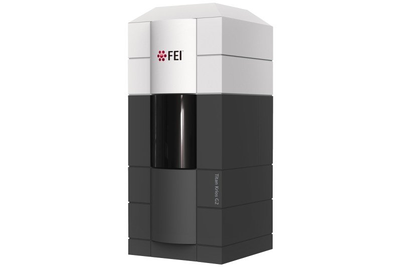
I am an FEI Titan Krios cryo-electron microscope with a Gatan K2 Summit detector. But most people will probably just call me the cryoEM.
How big are you?
I am 15 feet tall and I weigh 17,000 pounds.
Where will you live?
On the first floor of the Rockefeller Research Laboratories building in room 118.
Where are you now and will when you arrive at MSK?
I’m currently resting in a storage facility while they prepare the room for me — there’s a lot of retrofitting and redesign that needs to happen. I’m scheduled to arrive by the end of the year.
What is the difference between you and other electron microscopes?
I’m similar in that I shoot beams of high-speed electrons at a sample to generate an image that can be captured and projected onto a computer screen.
The main difference between these older microscopes and me is that I have a much more powerful electron detector on the receiving end, which allows me to capture details at a much higher resolution. It’s a lot like the burst mode function on your iPhone, in that it allows me to take many quick pictures during each exposure, which I then string together into a movie. By correcting for shifts in orientation of the sample between each frame, I can assemble a final image that is much crisper.
People say you will revolutionize biology by eventually replacing x-ray crystallography. What makes you better?
The main advantage is that you don’t have to first crystallize, or make a crystal out of, a sample. This is a very time-consuming step, and it doesn’t always work. Many proteins do not form crystals, but often you don’t know that until you’ve spent years trying to crystallize one.
Not having to make crystals also means that I can visualize a complete protein complex in its entirety, rather than just the one subunit that happens to be the right size and shape to form a crystal.
The other big advantage is that you need a much smaller sample to analyze. With x-ray crystallography, you need tens of milligrams of protein. For what I do, you need 1,000 times less. If I have a good sample, I can determine a structure within a week or two — compared to, in many cases, several years with x-ray crystallography.
What does the “cryo” part of your name mean?
That just means that the samples I shoot electrons at are frozen first; this helps keep them from vaporizing and also holds them relatively still.
What molecules are you most excited about sampling?
There are several. The mTOR kinases, which control cell growth and play a role in many cancers, are one large group of proteins. There are also several proteins involved in sensing and repairing DNA damage, such as BRCA2, ATM, and ATR that I’m eager to check out. Another is the protein PI3 kinase that is mutated in some breast cancers.
I am particularly excited about sampling membrane proteins, such as ion channels, since these proteins are very difficult to crystallize, and so are not well suited to x-ray crystallography.
Who will you be working with at MSK?
I will be collaborating closely with several research teams at the Sloan Kettering Institute, including those of Nikola Pavletich, Stephen Long, and Christopher Lima. With Dr. Pavletich, I’ll be working on the structure of oncogenes and tumor suppressor proteins. With Dr. Long, I’ll be focused on membrane proteins, and with Dr. Lima, the proteins involved in RNA processing. But I’m sure many other investigators will be eager to have me test their samples, too.
What do you say to those who think you look like a giant refrigerator?
Looks can be deceiving. They are probably speaking of my squarish container. This serves a clear purpose. Because the images I produce rely on a steady beam of electrons, it’s crucial that vibrations from the surrounding room be cancelled out. My square shell helps me do that. But I have a lot more interesting things inside, including a gun that fires electrons and powerful magnets that focus the electron beams. I am not a refrigerator.
Where do you see yourself in five years?
I suppose I will still be hard at work providing high-resolution structures of important biomolecules. By that time, maybe MSK will have acquired another cryoEM microscope; I wouldn’t mind some company.
This story originally appeared in the September 2016 issue of MSKNews.