
Getting it over the finish line was a labor of love — and now, more than five years after her death, the lab of former Sloan Kettering Institute Developmental Biology Chair Kathryn Anderson, PhD, is publishing its final study.
The work reveals how a critical signaling molecule called WNT guides cells in the embryo from a state of high flexibility — known as “plasticity” — toward distinct, specialized identities. The findings have implications not just for understanding developmental processes but for cancer biology as well. And it showcases how Dr. Anderson’s colleagues have worked for years to honor her legacy by ensuring that her lab’s final contributions to science could be realized.
Dr. Anderson was “an extraordinary scientist who made landmark discoveries at every stage of her career,” her obituary in the journal Cell noted. She joined Memorial Sloan Kettering Cancer Center (MSK) in 1996, ultimately serving as program chair until her death in November 2020. One of her lab’s overarching focuses was early mammalian development: how the highly plastic cells of the early embryo are directed to take on specialized identities needed to form the various organs and tissues of the body.
It was this line of inquiry that gave rise to the new paper, which was published July 1 Developmental Cell.
An intriguing mutation
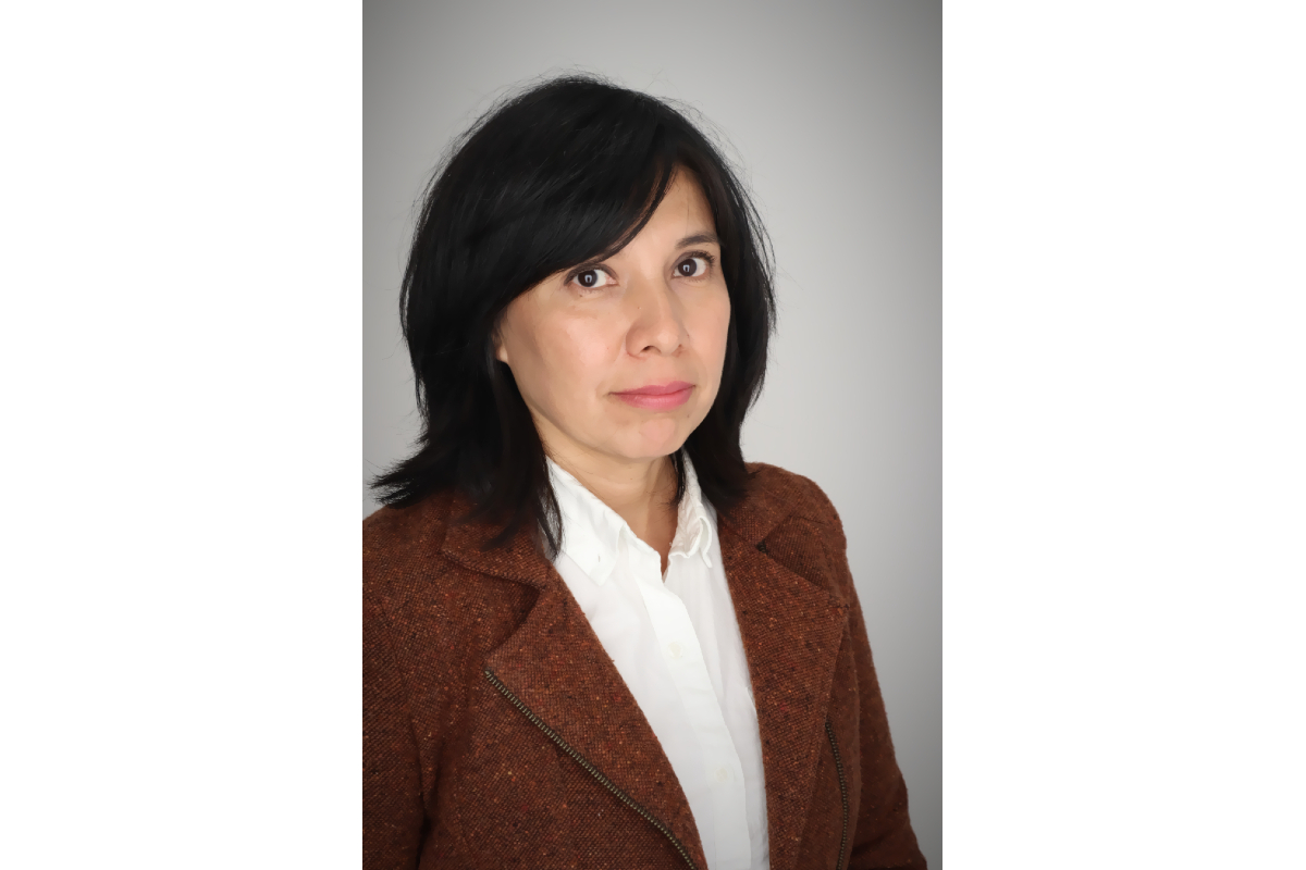
The project began about a decade ago, when Rocio Hernández-Martínez, PhD, then a postdoctoral researcher in Anderson’s lab, helped develop a mouse model with a striking abnormality. Using techniques to introduce genetic mutations, the team found that embryos missing two related genes — Axin1 and Axin2 — developed in a dramatically abnormal way.
Under normal circumstances, those genes act like a dimmer switch, keeping a key molecular signal called WNT from getting too strong. Without them, WNT gets stuck in the “on” position. The resulting embryos could only make a limited range of tissues — and were missing the cells that would normally give rise to the heart, head, and other front-of-body structures.
“This was a very interesting defect and so I adopted this project as my baby,” recalls Dr. Hernández-Martínez, who is now a senior scientist in the lab of Licia Selleri, MD, PhD, at the University of California, San Francisco, where she continues to study the genes and regulatory networks that shape embryonic development.
WNT’s multiple roles
For the current paper, the MSK team developed precise genetic tools that let them turn off the Axin genes specifically in the epiblast — a thin layer of highly plastic cells that serves as the starting point for essentially every tissue in the body. They paired those tools with single-cell sequencing to read gene activity in individual cells and track how cells communicate with one another. Together, these approaches allowed them to map out, in striking detail, how WNT shapes the earliest decisions a cell makes about what it will become.
What they found was that WNT doesn’t just do one thing, it acts in steps. First, it gives epiblast cells a push, nudging them out of their initial state — in which they could theoretically become any cell type — and starting them down the path toward becoming muscles, bones, organs, and connective tissue.
But the specific type of cell depends on a second layer of instruction, for which WNT is also critical.
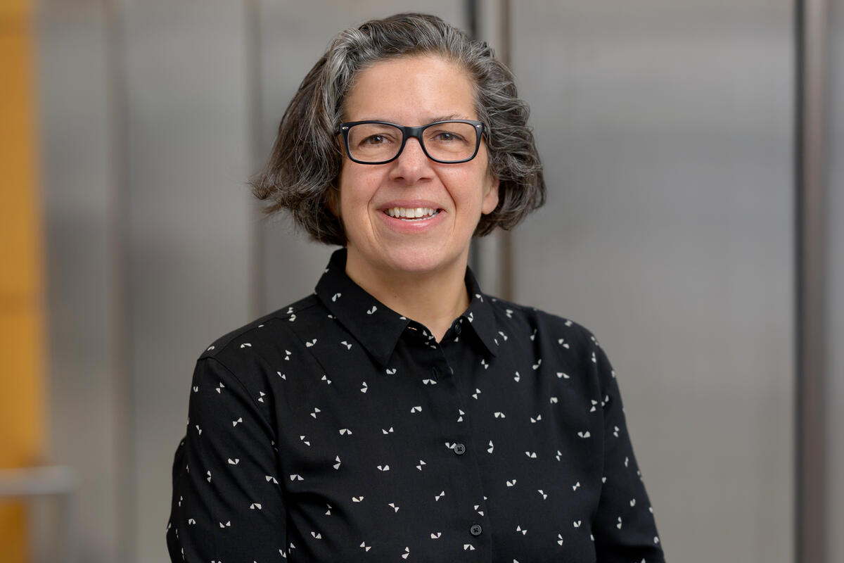
“WNT has multiple roles,” explains Anna-Katerina Hadjantonakis, PhD, a developmental biologist at MSK who oversaw the research after Dr. Anderson’s death. “It’s pushing cells from one state to the other initially, and then it’s integrating two distinct molecular signals across a spatial landscape to define the final outcome.”
Which of these signals a cell receives is based on its location, determining what identity it acquires and where in the developing embryo it ends up. Cells that encounter high levels of a molecular signal called BMP get directed toward the back of the body. Cells that encounter a different signal, called NODAL, are directed toward the front.
BMP and NODAL run in opposite directions across the developing embryo in a gradient — as one grows stronger, the other recedes. Each cell reads where it sits on that spectrum based on its location — like a topographic map — and combines that information with the WNT signal to determine its ultimate identity.
“BMP with WNT gives you one outcome; NODAL with WNT gives you another,” says Dr. Hadjantonakis, who succeeded Dr. Anderson as chair of the Developmental Biology Program. “And what makes that surprising is that BMP and NODAL belong to the same family of signaling molecules: TGF-beta. They’re related, but they drive cells in opposite directions.”
Implications for cancer metastasis
The findings also carry implications for one of cancer’s most dangerous traits: its ability to spread to other parts of the body, a process known as metastasis.
For a tumor to spread, cancer cells must first do something that cells can’t normally do — break away from neighboring cells and travel. The molecular program that enables this movement is the same one the study examines in the embryo, a process called epithelial-to-mesenchymal transition, or EMT.
In the early embryo, this freedom to move is by design — it’s how cells get where they need to go to build a body. In cancer, it marks the final and most aggressive stage of disease — as many as 9 in 10 cancer deaths are attributed to metastasis, making it a priority for researchers to better understand.
TGF-beta — the family of signaling molecules that includes both BMP and NODAL — is known to play a central role in driving EMT in cancer. But the new study suggests that treating TGF-beta as a single type of signal may be too simple. BMP and NODAL are both members of that family, yet they work through different molecular machinery and push cells in different directions. The outcome depends not just on whether TGF-beta is active, but on which TGF-beta is active.
“Not all TGF-betas are the same,” Dr. Hadjantonakis says. “BMP with WNT gives you one outcome. NODAL with WNT gives you another.”
For cancer research, that distinction may turn out to matter. Understanding the subtleties of how these signals interact in the developing embryo could help illuminate how they work in cancer — and point to new opportunities to slow or stop cancer’s spread.
Finishing what Dr. Anderson started
The science, however, almost never made it to publication.
First, Dr. Anderson fell ill and was overseeing her lab remotely. Then the COVID-19 pandemic hit and research across the institution slowed considerably as researchers navigated new restrictions to keep essential experiments going.
“We didn’t realize how sick she was because we were in lockdown,” Dr. Hadjantonakis recalls. “And then one day we learned that she’d passed.”
After her death, Dr. Anderson’s remaining lab members transitioned to new labs across the institution. Dr. Hernández-Martínez joined Dr. Hadjantonakis’ lab, where she spent two more years advancing the WNT project before taking a position at UCSF.
“Dr. Anderson was a wonderful mentor,” Dr. Hernández-Martínez recalls. “She used to come to the lab every morning and ask, ‘What are you doing? Can we talk about your project?’ She was always curious, always thinking about the science. Completing this project was the best way to honor her.”
But with Dr. Hernández-Martínez starting a new chapter of her career and no dedicated funding, the project became a shared responsibility — squeezed in around other work.
“It was everyone’s side project, but we were determined to see it through,” Dr. Hadjantonakis adds. Two members of the Hadjantonakis Lab stepped in to conduct further experiments to help steer the work to publication: senior research scientist Sonja Nowotschin, PhD, and senior research assistant Ying-Yi Kuo, MS. A collaboration with the lab of Bertie Göttgens, D.Phil, at the Cambridge Stem Cell Institute in the U.K. provided essential genomics expertise.
“It would have been such a shame just to have to drop it,” Dr. Hernández-Martínez says. “It’s such a beautiful science story.”
For Dr. Hadjantonakis, shepherding the project through to publication was both a scientific commitment and a personal one.
“In part it was a way of honoring a dear friend and colleague, but a way of also respecting that Kathryn was pursuing really fundamental questions about WNT’s role in directing cell identity in the embryo as a baseline for how it functions in other contexts,” she says.
The work also opens new questions about how a WNT signal integrates with BMP and NODAL signals at the molecular level.
Ultimately, in both a developing embryo and a growing tumor, cells are surrounded by competing signals — all talking at once — on an evolving 3D tissue landscape. The fundamental question is how a cell listens — filtering out some signals, acting on others, and arriving at a decision. In a healthy embryo, that process is exquisitely ordered. In cancer, it is more chaotic. Years in the making, this study brings us a step closer to understanding both, the researchers say.
Additional authors, funding, and disclosures
Additional authors on the study include Luke T.G. Harland, Bart Theeuwes, and Elizabeth Lacy.
The study was supported by grants from the National Institutes of Health (R01HD094868, R01DK127821), the Wellcome Trust (BI20-689, 226309/Z/22/Z), the Pew Latin American Fellows Program, and MSK’s Core Grant from the National Cancer Institute (P30CA008748).
Dr. Hadjantonakis is a member of the advisory board of Developmental Cell.
Read the study: “AXIN1 and AXIN2 regulate the WNT-signaling landscape to promote distinct mesoderm programs,” Developmental Cell. DOI: 10.1016/j.devcel.2026.06.004