T cells are an elite fighting force of the immune system, seeking out and destroying diseased cells. But in a prolonged campaign against a chronic condition — like a viral infection, or cancer — the body needs a steady supply of these killer troops. Where and how these killer troops are generated has been a mystery.
That led a team of scientists from Memorial Sloan Kettering Cancer Center (MSK) and Weill Cornell Medicine to dig deeper. They found that a small subset of T cells, called stem T cells, are responsible for making new T cells and for continuously replenishing them in chronic disease. Importantly, these rare stem T cells express a protein called LEF1.
The team’s findings in laboratory models, published July 1 in Cell, showed that focusing on this population of LEF1-positive T cells is key. Boosting LEF1-positive cells overcame T cell “exhaustion” in the case of chronic infection. And removing them was successful in reining in overactive immune cells in the case of type 1 diabetes, an autoimmune disease.
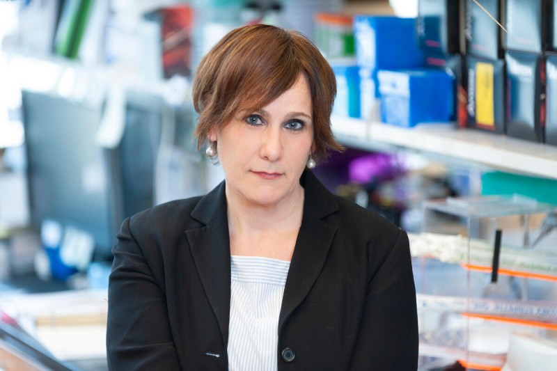
The research also has the potential to inform future cancer treatments, says senior author Andrea Schietinger, PhD, a cancer immunologist in MSK’s Sloan Kettering Institute.
“Although it was not part of this study, cancer is a chronic disease where T cells lose their capacity to fight cancer cells over time,” she says. “So that’s what we’re looking at next.”
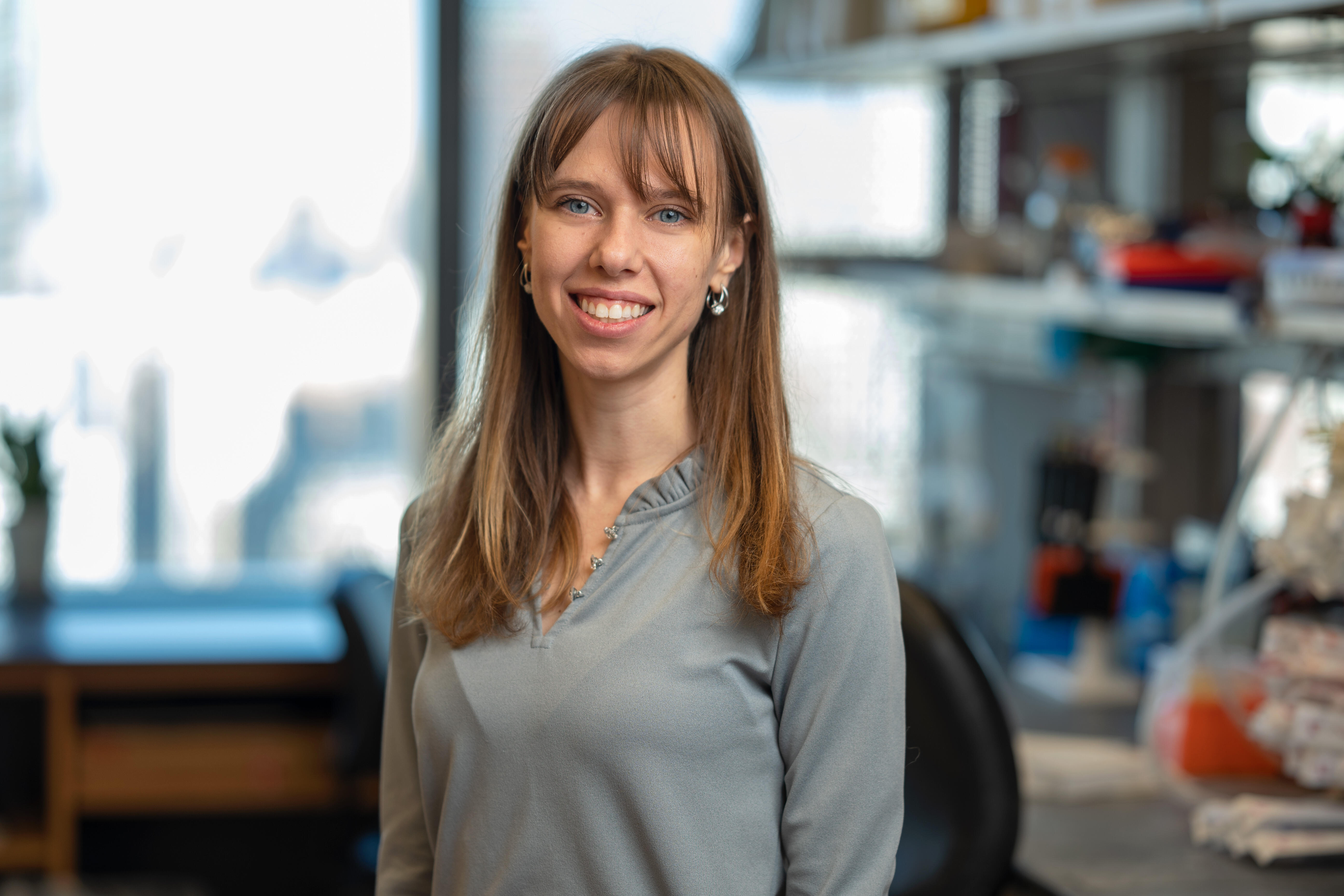
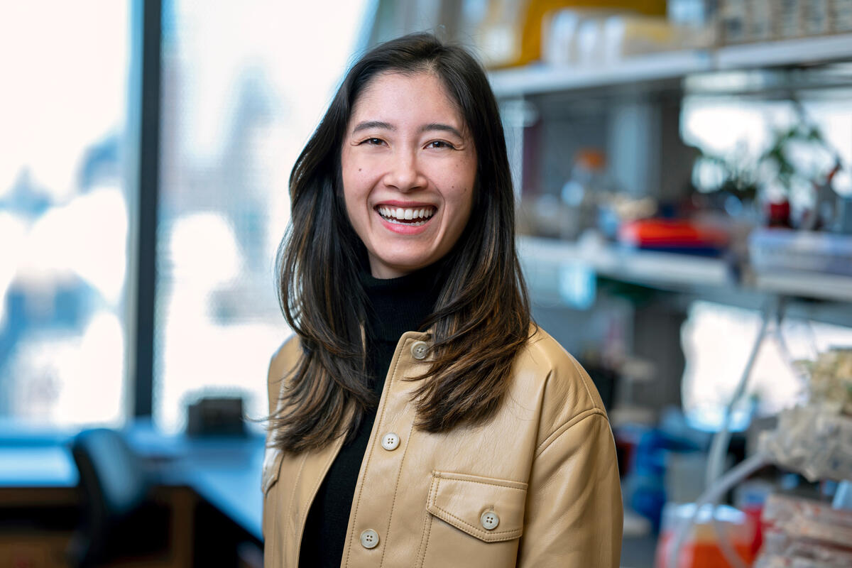
The study was led by co-first authors Svetlana Miakicheva and Katrina Hawley, PhD, both members of the Schietinger Lab at MSK, and by Paul Zumbo, a senior staff scientist in the lab of co-corresponding author Doron Betel, PhD, at Weill Cornell Medicine.
Showing LEF1 Is Critical for Stem T Cells
To prove that LEF1 wasn’t just a marker of stem cells (or “stemness”), but a central player, the researchers used CRISPR gene editing to delete the LEF1 gene from these rare stem T cells in their mouse models.
The results were striking. Without LEF1, stem T cells lost their ability to persist and self-renew.
In the autoimmune diabetes model, mice whose T cells lacked LEF1 were significantly protected from developing the disease because the disease-causing T cells could no longer sustain themselves and destroy insulin-producing cells in the pancreas.
Meanwhile, going in the other direction proved equally revealing. When the researchers boosted LEF1 levels, more stem T cells formed — and fewer cells reached the terminal, “burned out” stage in the viral infection model.
“Our study shows that LEF1 is key to T cell stemness and persistence,” Dr. Schietinger says. “Turn it up, and you get more stem cells. Remove it, and the stem cell pool disappears. Which of those is desirable depends on the disease context.”
Different Diseases, Same Biological Playbook
One of the most surprising findings came when the team compared stem T cells from the two diseases side by side: autoimmune diabetes and chronic infection with a virus called lymphocytic choriomeningitis — a well-established model for studying chronic viral infection in mice.
On the surface, these conditions couldn’t be more different. In autoimmune diabetes, T cells are highly active and aggressive — destroying healthy insulin-producing cells in the pancreas. In chronic viral infection, T cells become functionally “exhausted,” burning out over time and allowing the virus to persist.
And yet, when the researchers mapped the molecular profiles of both cell types using a computational visualization technique, the two stem T cell populations clustered together as a single group — essentially indistinguishable from one another. This suggests that LEF1-driven stemness isn’t a disease-specific quirk, but rather a shared feature of how the immune system sustains itself under chronic stress; the team found 117 genes across both diseases that share the same pattern of being switched on or off.
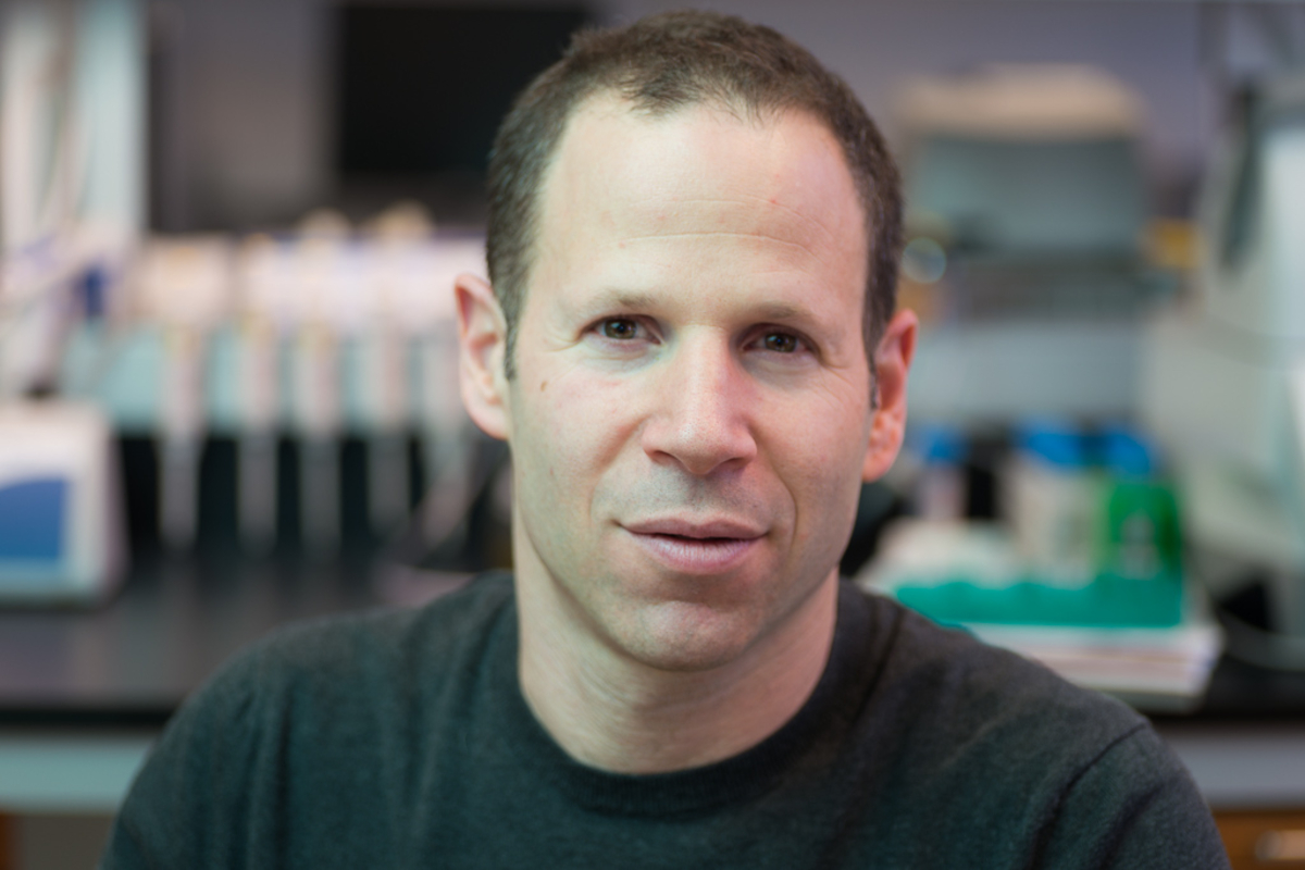
“This points to a common underlying mechanism of stem T cell state, driven by LEF1, that is shared across these two very different diseases,” says Dr. Betel, whose lab performed the sophisticated computational analyses needed for the project. “LEF1 drives a fundamental mechanism by which the immune system sustains stem T cells during chronic infection, as well as drives autoimmune conditions, rather than being unique to a particular disease. This opens the possibility to new therapeutic strategies for a broad range of immune related conditions.”
And how do these stem T cells sustain themselves?
The authors were surprised to find that many genes and pathways employed by stem T cells matched those of embryonic and adult stem cells, which are found throughout our tissues, including the skin, intestine, muscle, and bone marrow.
Location, Location, Location
Similar to stem cells in the gut or the bone marrow — which depend on specialized environments called “niches” — the location of immune stem T cells matters. Each T cell population expressed different molecular “address labels” directing them to distinct locations within lymph nodes and tissues. In collaboration with the lab of MSK physician-scientist Ivan Maillard, MD, PhD, the authors disrupted those location signals — either by blocking proteins called integrins or interfering with a pathway called Notch signaling — and strikingly, the stem T cell pool collapsed.
“Stemness isn’t just about what’s inside the cell,” Dr. Schietinger says. “It’s also about where the cell lives and what signals it receives from its environment.”
Moving the Findings From the Bench Toward the Bedside
For Dr. Schietinger and her colleagues, the findings also underscore the importance of doing research into fundamental human biology, which is often called “basic science.” The idea is that by working to understand how T cells replenish themselves, new therapeutic strategies may emerge.
“We’ve identified what we believe is a fundamental mechanism, one that the immune system uses broadly to sustain itself in chronic disease,” Dr. Schietinger says. “That’s the kind of finding that can open up entirely new directions for treatment.”
In this case, disrupting stem T cells could potentially block them from attacking an individual’s own tissues in the case of autoimmune disorders. Or, alternatively, in the case of viral infections — or cancer — the pool of stem T cells could be boosted, helping the immune system to maintain a durable fighting force.
“This work demonstrates the power of multidisciplinary collaboration where well-designed disease models, cutting edge experiments, and advanced computational analysis are brought together to address important scientific questions,” Dr. Betel says.
The work also dovetails with a larger research effort at MSK known as the Marie-Josée and Henry R. Kravis Cancer Ecosystems Project, which is supported by a generous philanthropic gift. Its goal is to understand cancer not just as a disease of mutated genes, but as one in which cancer cells communicate with the immune cells and tissues around them to support a tumor’s growth.
“Understanding how T cells sustain themselves — and how their environment shapes them — is fundamental to understanding cancer,” Dr. Schietinger says. “Engineering niches and locations where cancer-fighting stem T cells can form and maintain themselves is at the heart of our research now.”
Additional Authors, Funding, and Disclosures
Additional authors of the study include Sofia Gearty, Simon Grassmann, Brianna Naizir, Edison Chiu, Sandra Carson, Michael Kissner, Mark Owyong, Yunlin Zhang, Robert Washburn, Joseph Sun, Ronan Chaligne, and Steven Reiner.
The study was supported by the National Institutes of Health (R01AI173249, F31DK141119, F30 DK122691, R01CA279268, R01AI091627, CA278976), the Breakthrough T1D foundation (SRA-2023-1410-S-B), a Lloyd J. Old STAR Award of the Cancer Research Institute, an MSK Basic Research Innovation Award (BRIA), and Hearst Foundation and MSK–Cancer Research Institute Fellowships. The research also relied on the MSK Integrated Genomics Operation, Flow Cytometry Core, and Single Cell Analytic Innovation Lab (SAIL), which are supported by MSK’s National Cancer Institute Cancer Center Support Grant (P30 CA08748). The work received additional support from Cycle for Survival, the Marie-Josée and Henry R. Kravis Center for Molecular Oncology, the Weill Cornell Medicine Core Laboratories Center, Columbia University Stem Cell Initiative Flow Cytometry Core Facility, and the NIH Tetramer Core Facility.
The authors declare no competing interests.
Read the study: “LEF1 and niche factors determine T cell stemness across chronic diseases,” Cell. DOI: 10.1016/j.cell.2026.06.022