Computational Image Analysis Laboratory
Overview

Binsheng Zhao
Director, Computational Imaging Analysis Lab; Member, Radiology

Lawrence H. Schwartz
Chair, Department of Radiology
The Computational Image Analysis Laboratory is dedicated to advancing cancer research and clinical practice by analyzing cancer imaging phenotypes and integrating genotypes using state-of-the-art image processing, radiomics, and deep learning techniques, with the goal of enhancing clinical decision-making in personalized precision oncology. We focus on developing and automating novel quantitative imaging biomarkers derived from CT, MRI, and PET modalities for a wide range of clinical applications, including (but not limited to) cancer surveillance, diagnosis, prognosis, and early response assessment across all solid tumors.
Key projects
- Response Assessment Platform Integrated with Digital Volumetric Tumor Segmentation (RAPID-VTS)
- Radiomics pipeline developed for analyzing diverse imaging phenotypes
- Clinical validation of volumetry and kinetic modeling in tumor response assessment
- Automated detection and segmentation of solid tumors on CT images
- Automation of RECIST (AutoRECIST)
- Baseline response prediction and early treatment evaluation for oncology clinical trials
- Prostate vision-language model (VLM) to automatically generate radiology reports
- Automated surveillance, diagnosis and response assessment for kidney cancer
- AI-driven prediction of post-surgical recurrence in lung cancer
- Exploring and improving the reproducibility of radiomic features and signatures
- QA programs to ensure the accurate and reproducible quantitative imaging biomarkers
- Data sharing efforts
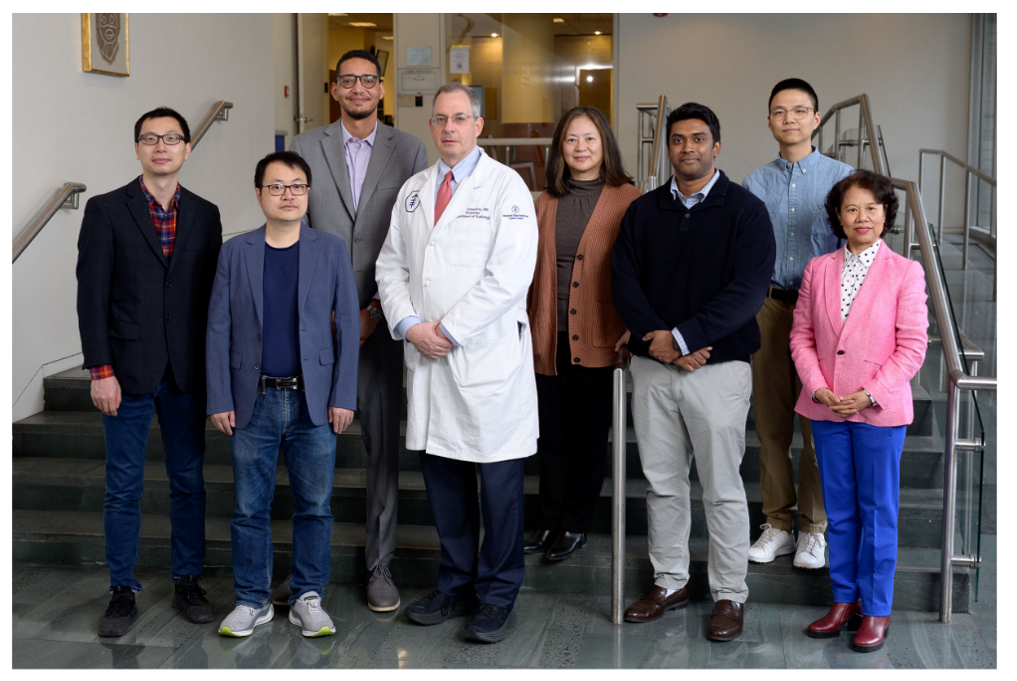
People


Director, Computational Imaging Analysis Lab; Member, Radiology
Chair, Department of Radiology
Members
Senior Research Scientist
Senior Research Scientist
Senior Research Scientist
Research Scholar
Visiting Investigator
Associate Lab Member
Bioinformatics Software Engineer
Visiting Investigator