Over the last few decades, there has been a dramatic increase in the availability and application of tests that use ionizing radiation to take images of the body. These tests include computed tomography, or CT, scans and nuclear medicine exams that use small amounts of radioactive material such as nuclear stress tests, bone scans, and PET scans.
Physicians use these tests to help diagnose disease, as well as to make treatment decisions and monitor a patient’s response to therapy. However, some media reports have suggested that doctors may be overusing these types of tests, especially CT scans, and exposing patients to unneeded radiation.
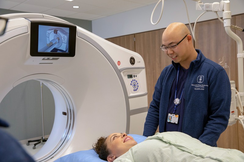
We asked Memorial Sloan Kettering Cancer Center (MSK) medical physicist Lawrence Dauer, PhD, to explain what you should know about the safety of these scans, how and why doctors use them, and what’s being done to address patients’ concerns.
Do tests like CT scans increase my risk of getting cancer?
Advances in technology and medical physics practice have helped to reduce the dose of radiation that patients receive from tests like CT. Radiation exposure is measured in units called millisieverts, or mSv. The effective dose from a diagnostic CT procedure depends on the area of the body being imaged as well as the size of the patient and typically ranges from about 1 to 10 mSv.
To put that in perspective, radiation protection experts estimate that the average person in the U.S. receives about 3 mSv per year from natural background radiation, which includes cosmic radiation from outer space.
Risk evaluations currently rely on information from large populations who have received much higher doses of radiation, such as patients undergoing radiation therapy or atomic bomb survivors. This information is extracted and applied to an individual getting a CT scan, which yields far less radiation exposure and much less potential theoretical risk than those examples.
For the average person, a CT scan may be associated with a very small potential theoretical risk — perhaps less than about .05 percent, or less than one in 2,000 — of possibly developing a future cancer. If we’re trying to figure out whether you have cancer or how best we should treat an existing cancer, such a small potential theoretical risk is far outweighed by the clinical benefit, which could save your life.
How do doctors decide whether a CT scan is necessary?
MSK physicians rely on two fundamental principles to protect patients when it comes to using imaging tests that employ radiation. The first is justification. We don’t order a test unless it’s justified, meaning that the clinical benefit outweighs any associated risk. Once we’ve ordered the test, we implement the second principle, which is optimization — obtaining the best image we can with the lowest dose of radiation in order to reduce any associated potential future risk.
MSK’s collaborative team of oncologists, radiologists, nuclear medicine physicians, medical physicists, and radiation safety staff work hard to ensure you receive the lowest possible amount of radiation exposure from each imaging test without compromising image quality.
We want to give our physicians the very best tools and information to help them make informed diagnostic and treatment recommendations and engage with patients in shared decision-making about their care.
Can getting too many CT scans be dangerous?
Not if they are justified and optimized. If those two principles are in place, then the benefit of having the test is going to far outweigh the potential risk. If you have symptoms of an illness, the information that can be obtained from a scan is usually justified. However, if we are talking about using imaging for screening purposes and you have no symptoms, we need to carefully evaluate whether you really need that test.
For example, CT screening for lung cancer has been greatly debated. It’s not right for everyone, so we have to ask, “Are you a smoker? Are you of a certain age?” If you fall into a high-risk category, then we can say that CT screening is a justified and appropriate diagnostic tool for you.
Screening CT renders a much lower dose of radiation compared with the kind of CT scans we use to look at a suspected lung cancer in someone with symptoms or to make treatment decisions. Because those scans require a greater amount of information about the image, the dose is appropriately higher.
How many CT scans are safe in a month or a year? How many is too many?
If the scans are justified for diagnosis or treatment evaluation, there is no set number. For example, even two or more scans in a week or four in a month could be appropriate depending on your particular disease management. Again, for justified imaging scans the risks are low compared with the clinical benefits. From a safety standpoint, there is no set time interval required between scans.
Are certain organs more sensitive to radiation exposure than others?
There are differences in organ sensitivity to radiation. For example, the breasts, thyroid, lungs, and bone marrow are more sensitive to radiation because the cells in those areas divide rapidly. Less-sensitive organs include the brain, where cells don’t divide as quickly.
Our assessments — such as that nominal .05 percent potential risk for a typical CT — factor in this wide range of organ sensitivity. We use mathematical models and other simulation tools to assess where radiation travels in the body and how it interacts with organs in order to get a better picture of dose distribution, measure the risk to the site we are imaging, and reduce the amount of radiation that scatters to areas outside of the site being imaged.
Are children more sensitive to radiation than adults?
Yes, because their cells divide rapidly. Size and weight also matter when determining the appropriate dose and minimizing radiation scatter to surrounding organs during imaging. When imaging children, we emphasize justification and implement child-size protocols to help minimize their overall exposure.
What is being done to understand and address patient concerns about imaging risks?
Our research has shown that most patients with symptoms want their physicians to help them figure out what’s wrong and choose the best treatment. But once treatment is over, survivors may have more reservations about being scanned and are concerned about whether follow-up imaging might raise their risk of recurrence.
We’ve also learned from national data that some patients may refuse imaging exams because they are afraid of the risks associated with them.
We can’t ignore the fact that people feel this way. We need to listen to patients and find better ways to communicate with them about the benefits and risks of imaging at the time of diagnosis and treatment as well as after treatment.
In certain patients, it makes sense to check for signs of recurrence with periodic scans. Other patients can be imaged less often. In all cases, MSK ensures that there is evidence to support a change in a patient’s management based on the imaging tests being ordered. That’s what you need for proper justification.
Can I protect my body from radiation during or after a CT scan?
The use of shielding during CT scans is not recommended because the shields can significantly affect image quality and reduce the ability of physicians to properly diagnose disease or evaluate treatment outcomes. In addition, CT scans do not result in making any part of your body radioactive. The x-rays used to make the images pass through the body. Therefore, there are no actions required after a CT scan.
Should I get a full-body CT scan to detect diseases like cancer early?
If you have symptoms, an illness, or a particular predisposition for a disease, and a physician prescribes a full-body CT scan, then the information that can be obtained from such a scan is usually justified. However, as noted earlier, the use of imaging for screening of healthy people or those without symptoms is otherwise not recommended since the benefit may not outweigh the associated potential risks.
Should physicians consider using alternative imaging methods such as ultrasound or MRI?
Yes, we do consider alternatives for certain cases when appropriate. However, not all imaging gives us the same information. We can’t replace all CTs with MRIs for example, but when it makes sense, and it’s justified, we should.
This story was originally posted in 2015 and has been updated.