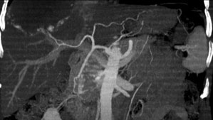
At Memorial Sloan Kettering, doctors are investigating a new technique to view the blood vessels that feed cancers of the liver. Computed tomographic (CT) angiography, an imaging technique frequently indicated for vascular disease, may also provide clear views of the vessels that feed hepatobiliary tumors.

|
| This example of a CT angiographic image shows the arteries feeding a large but benign liver mass (upper left). |
Administered on an inpatient or outpatient basis, CT angiography is usually performed in conjunction with a standard abdominal CT scan; together, the tests take about 10 minutes. Like a standard abdominal scan, CT angiography begins with the injection of a contrasting agent (a dye) into a vein in the arm. Although the dye highlights the liver, pancreas, and tumors on the results of the scan, the blood vessels that supply the tumors may appear poorly defined. In CT angiography, however, radiologists inject the dye at a much faster rate, then take a series of pictures, at rapidly paced intervals, of the blood as it flows to the affected area. A computer processes these pictures into 3D images that show surgeons the relationship of important vessels to cancerous growths and help them delineate any normal variants, both of which are vital to the development of a safe and successful surgical plan.
Studies comparing the utility of CT angiography with that of more conventional diagnostic procedures are under way. Patients with kidney failure or known allergies to intravenous contrasting agents should not have the test, but otherwise, side effects and discomfort have been generally mild. CT angiography is minimally invasive (only a vein is punctured) and requires no sedation, while more conventional diagnostics like an angiogram, for example, involve the insertion of a catheter into the femoral artery and do require sedation.
We’re available 24 hours a day, 7 days a week