Convection enhanced delivery (CED), an innovative technique that directly infuses a therapeutic agent into a brain tumor through a cannula, bypassing the blood-brain barrier (BBB), is safe and feasible for treating diffuse intrinsic pontine glioma (DIPG), according to our experience to date in an ongoing phase I clinical trial at Memorial Sloan Kettering Cancer Center (MSK). However, despite the significant advantages with CED, numerous hurdles remain.
Together with co-author Umberto Tosi, MD at Weill Cornell Medicine, I recently published a review paper in Pharmaceutics to provide an update on our experience using CED to treat pediatric patients with DIPG. (1)
The experience has alleviated many of the concerns related to the safety and feasibility of using CED in children with DIPG. Median overall survival (OS) was 17.5 months, and the one-year survival rate was 58.5 percent for the first 25 patients in our trial. (2) A few children have survived more than three years post-treatment, providing glimmers of hope that CED may prove to be an essential tool for changing DIPG’s typically dire prognosis.
In our review paper, we also discuss the advantages, technical advancements taking place in the field, and the hurdles that will need to be overcome before demonstrating clinical benefit with CED. (1)
DIPG: The Challenging Landscape
Pediatric diffuse midline gliomas (DMGs) have universally poor prognoses. Despite representing only 10 to 20 percent of all pediatric central nervous system malignancies, they are the most prevalent cause of death due to brain cancer in children. (3), (4) DIPGs are a particularly aggressive, inoperable subtype of DMG, characterized by an infiltrative process in the brainstem. The incidence of DIPGs peaks at six to nine years of age, rarely occurring in adolescents and adults. Median survival after initial radiation therapy is 11 months. Overall survival (OS) is 30 percent at one year and 10 percent at two years, with only occasional reports of survival at five years. (5), (6) This poor prognosis has not changed for decades.
Patients usually present with a triad of worsening cranial nerve deficits, cerebellar symptoms, and long tract signs, and characteristic magnetic resonance imaging (MRI) that leads to diagnosis. (7) At present, radiation therapy is the only approach that improves progression-free survival (PFS), and only for a limited time.
DIPGs are astrocytomas that spread along fiber tracts. The thalamus and cerebellum are the most common sites. (8), (9), (10) It is believed that the spread involves cells that diverged early during tumor development and abandoned their niche. As a result, these cells are strikingly different from those that constitute the bulk of the tumor, implying a predisposition to drug resistance and disease recurrence. (11)
Challenges to Treating DIPG
There are numerous challenges to treating DIPG. Cytoreductive surgery is not possible given its highly infiltrative growth patterns and position within the pontine segment of the brain stem. Since biopsies are associated with high morbidity, (12) little tissue has been available, hindering molecular and genetic profile studies and the development of preclinical models. Recently, minimally-morbid stereotactic biopsies have helped establish such models, allowing for more research. (4), (12), (13), (14)
Conventional agents have shown little to no success. (6), (15), (16) Temozolomide (TMZ), either delivered concurrently or after initial radiotherapy, failed to achieve any survival benefit and resulted in significant toxicities. (15), (16) Results for coupling TMZ with cis-retinoic acid (17) or thalidomide (18) were similarly abysmal. Traditional chemotherapies have also failed to prolong survival and often resulted in significant hematological side effects. (19), (20), (21), (22), (23)
Targeted therapies have also shown poor responses and significant toxicities. Despite notable preclinical promise, the following agents failed: tamoxifen (an estrogen-receptor modifier), (24), (25) bevacizumab (a vascular endothelial growth factor-VEGF-inhibitor), (26) nimotuzumab (a monoclonal antibody inhibitor of epidermal growth factor receptor-EGFR), (27) gefitinib (a small molecule EGFR inhibitor), (28) erlotinib (an EGFR inhibitor), (29) and dasatinib (a BCR-Abl kinase and platelet-derived growth factor receptor A (PDGFRA) inhibitor). (30)
Immunoreactive agents such as monoclonal antibodies and chimeric antigen T cells (CAR T cells) are providing glimmers of preclinical promise. Their potential traces to their ability to target non-dividing cells that traditional chemotherapies do not address. (31) Most CAR T cell therapies are still in early phase clinical trials, so it’s too soon to conclude on their efficacy. (2), (32), (33) Further, their efficacy may be hindered by high levels of tumor heterogeneity with DIPG cells, and treatment-associated inflammation can result, rarely, in hydrocephalus, which is a lethal complication. (34)
One of the main issues with treating DIPG with systemic therapies is the inaccessibility of the tumor due to the tight endothelial junctions of the BBB that impede drug penetration. (35) Traditionally, drug dosage has been increased to try to improve tumor penetration to therapeutic levels, but dose-limiting toxicities have ensued. Hundreds of clinical trials in DIPG have not met their primary endpoints despite preclinical promise, because systemic toxicity has impeded dose escalation to therapeutic levels. (36)
Other methods to get past the BBB include disruption via chemical means or through ultrasound, (37) intra-arterial delivery, the use of carriers, and local delivery. Lack of spatial sensitivity, lack of tumor specificity, or significant toxicity has made disruption methods problematic. (38) Intra-arterial therapy does not directly circumvent BBB impermeability, and the pontine location of DIPG and lack of a single arterial feeder weaken the case for this method. (39) Carriers developed so far lack tumor-targeting properties that would localize their distribution within the DIPG tumor and cause significant central nervous system toxicities. (40)
Convection Enhanced Delivery (CED) for DIPG
CED delivers a therapeutic agent directly into a brain tumor through a cannula implanted in a surgical procedure. The technique has been developed for at least two decades, first in preclinical animal models, (41), (42) and more recently in early phase clinical trials for treating primary or recurrent high-grade gliomas. (36), (42), (43), (44)
CED has numerous additional advantages compared to systemic therapies. It relies on a pressure gradient to deliver the infusate, enabling continuous delivery at an almost-constant concentration and even permeation of homogenous tissue.
DIPG is a logical tumor for using CED, since it is constrained within a limited anatomical compartment, making drug distribution more easily achievable, in theory. To date, some clinical studies have demonstrated safety and feasibility of the approach with various agents. However, large variability in technique, infusate, and hardware in these studies, made it difficult to draw unifying conclusions about the efficacy of CED. (45), (46), (47), (48), (49), (50)
Phase I Clinical Trial: Experience to Date
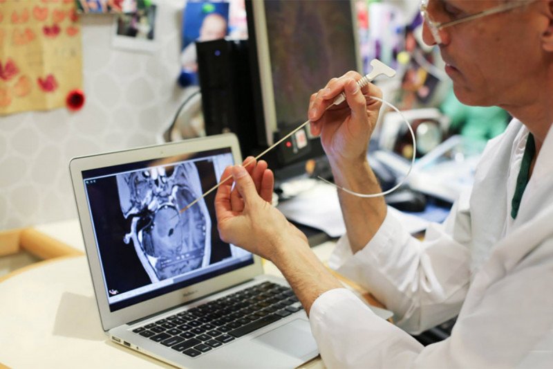
Our phase I clinical trial NCT01502917 is the first to treat pediatric patients with DIPG with an iterative dose, volume, and rate escalation design. The primary objective is to evaluate the safety of CED in children with DIPG and to define safe infusion parameters. We are monitoring patients using both MRI and positron emission tomography (PET).
Children with a clinical and radiographic diagnosis of nonprogressive DIPG who have received standard radiation therapy are eligible to enroll. Study participants are infused with the radiolabeled monoclonal antibody Omburtamab (y-mAbs Therapeutics; New York, NY, USA) between four and 14 weeks after the initial radiation therapy. Omburtamab targets the membrane-bound protein CD276 (B7-H3), an immune modulator part of the B7 superfamily overexpressed in DIPG and other pediatric cancers of the central nervous system. (51), (52), (53)
Our trial design relied heavily on treatment planning and novel technologies repurposed for CED. We obtained an investigational exemption to use a semi-flexible catheter (BrainLab, Munich, Germany) that is inserted using magnetic resonance (MR) guidance (Clear Point Neuro, Irvine, CA, USA). Infusions of Omburtamab were administered at the bedside in an intensive care unit, with volumes ranging from 240 μL to 4540 μL, for a prescribed dose from 0.25 mCi to 4.00 mCi.
To date, the average volume of distribution to volume of infusion (Vd/Vi) ratio has been 3.4 ± 1.2, which means we are achieving distribution volumes of about 12 mL for the highest published single infusion dose level of 4,000 μL. The average lesion-to-whole body absorbed dose ratio is more than 1,000, indicating that CED is attaining very high intralesional doses with negligible systemic exposure. This is the inverse of what happens with systemic drug delivery, where only a fraction of the drug reaches the tumor. (2) Since our initial experience, we have successfully demonstrated the safety of CED in treating children with DIPG with volumes of infusions greater than 8,000 μL, with Vd up to 35 mL, allowing for coverage of most post-radiation tumor volumes.
As of May 2020, no grade IV treatment-related adverse event has occurred. Grade III toxicities have included left-side muscle weakness, ataxia, and dysarthria in one patient and generalized muscle weakness in another patient, adding to the previously reported transient hemiparesis and one skin infection. (2)
In our first published report of results for 25 patients, median OS was 17.5 months, and the one-year survival rate was 58.5 percent, surpassing most cooperative group trials. Longer-term survival has been demonstrated in a few children so far — three have survived more than three years, and one has survived six years post-treatment — providing hope that CED may one day prove to lengthen survival. (2)
However, as with any phase I trial, it is impossible to make definitive conclusions about survival benefit or efficacy. There is notable variability in our study participants for the timing of treatments, tumor burden, prescribed dose, distribution volume, absorbed dose, and tumor coverage. Our trial is ongoing with an expanded cohort seeking to recruit 64 patients.
Optimizing CED for DIPG
In our clinical trial, we estimated Vd as a change in T2-weighted signal on the post-infusion magnetic resonance imaging (MRI) compared to baseline pretreatment MRI. We have observed a Vd/Vi ratio of about three in our patients treated so far, consistent with other preclinical and clinical research. (2), (43), (45), (54), (55), (56) However, the longitudinal behavior of distribution remains unknown.
There are a limited number of therapeutics that can be directly imaged with PET. However, recent developments in synthetic radiochemistry have expanded the library of compounds that can be transformed into therapeutic diagnostics. We have experience in creating [124I]- and [18F]- labeled agents. (57), (58), (59), (60), (61), (62), (63) The former has a half-life of 4.2 days, allowing for more accurate long-term tracking of drug behavior. The latter decays rapidly, with a half-life of 110 minutes, reducing patient exposure to ionizing radiation but making it a preferred isotope for patients requiring multiple doses.
There should be at least a complete overlap between Vt and Vd to achieve maximal tumor coverage with a therapeutic compound. Our estimates of drug-tumor intersect reveal a significant variance, ranging from 25 to 96 percent. Smaller tumor volume and greater infusion volume would both produce greater Vd and tumor intersect. Enhancing targeting to avoid longitudinal white matter tracts, pial or ependymal surfaces, and necrotic or cystic regions will also play a role in optimizing tumor coverage.
Future trials aimed at demonstrating a clinical benefit will need to measure the degree of tumor coverage to assess whether it impacts outcomes. Measuring and predicting tumor coverage may be improved with simulation software that employs tissue imaging characteristics and infusion parameters. (64), (65) Despite numerous hurdles to using them in the clinic, we have observed significant concordance between predicted and actual Vd since we adopted them.
Another issue pertains to the behavior of infusates. Since convection relies on establishing a pressure gradient between infusion front and brain parenchyma, infusates can fall into low-pressure wells, such as tumor cystic components or catheter tract via backflow. These issues are being addressed by new step catheters and new implantable devices with single or multiple catheters to allow for re-dosing or continuous infusions. (45), (55), (56), (66)
One of the main issues with implantable systems is that a second infusion in the same target is likely not beneficial, since it would occur in mainly necrotic tissue from the first infusion. In our experience, patients who have been re-dosed a second or third time with 124I-8H9 required new surgeries for each intervention and tolerated the procedures well.
The lack of accurate and non-invasive drug monitoring tools compounds these challenges. Except for radiolabeled agents such as 124I-8H9, the distribution volume and pharmacokinetics profile of most infused chemotherapeutics cannot be easily visualized.
Co-infused agents may be effective for estimating distribution at time zero, but they are not reliable long-term. (67) New theranostics are being developed for CED, based on modifications of small molecules, (62), (68), (69) or relying on larger carriers. (37), (58), (59), (71) These agents hold promise but have not been successfully translated to the clinic yet.
The Paradox of Using CED for DIPG
It remains unclear whether CED may be curative for DIPG. Recent studies have shown that up to one-third of patients had leptomeningeal disease spread, and one-fourth had disease outside the brainstem at the time of autopsy. (8)Clonal analysis of pontine and extra-pontine tumor samples has revealed that migration from the brainstem occurred early during disease progression, possibly before diagnosis and initial radiation therapy. (11)
CED holds promise for achieving regional disease control. Coupled with other innovative approaches, such as craniospinal radiation or intrathecal delivery of chemotherapeutics, CED may improve DIPG’s dire prognosis. (72)
The optimal agent or combination of agents to be administered via CED is still a matter of debate. In recent years, there has been a notable increase in CED-based clinical trials for DIPG beyond our NCT01502917 as follows: NCT00880061 is testing IL13-Pseudomonas toxin; NCT03566199 is testing a water-soluble version of the histone deacetylase complex inhibitor Panobinostat; and, NCT03178032 is investigating DNX-2401, an oncolytic virus.
Collaboration is Essential for Improving DIPG Outcomes
The rise of collaborative efforts aimed at improving the prognosis of DIPG has been one of the few positive notes in recent decades. Not-for-profit foundations have been sponsoring research since the 2000s, resulting in the foundation of the DIPG Collaborative and its DIPG Registry — a comprehensive database of clinical, radiological, pathologic, and molecular data.
Governmental funding agencies and private donors have also advanced DIPG research substantially. Overall, collective efforts have led to the development of promising preclinical models, the discovery of novel pathways, and the development of targeted therapeutics.
Together, we will continue pioneering innovative treatment approaches that show potential for improving DIPG’s abysmal prognosis.
Dr. Souweidane is a pediatric neurosurgeon at MSK, director of pediatric neurological surgery at Weill Cornell Medicine, and principal investigator for the clinical trial NCT01502917. Dr. Souweidane declares no conflict of interest, financial or otherwise.
The present study received no external funding. The authors are grateful to the pediatric neuro-oncology and neurosurgery departments of Weill Cornell Medicine and MSK for their help and contribution in carrying out NCT01502917, which has been sponsored by Y-mAbs since 2017. Omburtamab was licensed to Y-mAbs Therapeutics in 2015.
- Tosi U, Souweidane M. Convection Enhanced Delivery for Diffuse Intrinsic Pontine Glioma: Review of a Single Institution Experience. Pharmaceutics. 2020;12(7):E660.
- Souweidane MM, Kramer K, Pandit-Taskar N, et al. Convection-enhanced delivery for diffuse intrinsic pontine glioma: a single-centre, dose-escalation, phase 1 trial [published correction appears in Lancet Oncol. 2018 Aug;19(8):e382]. Lancet Oncol. 2018;19(8):1040–1050.
- Kebudi R, Cakir FB. Management of diffuse pontine gliomas in children: recent developments. Paediatr Drugs. 2013;15(5):351–362.
- Panditharatna E, Yaeger K, Kilburn LB, et al. Clinicopathology of diffuse intrinsic pontine glioma and its redefined genomic and epigenomic landscape. Cancer Genet. 2015;208(7–8):367–373.
- Grasso CS, Tang Y, Truffaux N, et al. Functionally defined therapeutic targets in diffuse intrinsic pontine glioma. Nat Med. 2015;21(7):827.
- Robison NJ, Kieran MW. Diffuse intrinsic pontine glioma: a reassessment. J Neurooncol. 2014;119(1):7–15.
- Schroeder KM, Hoeman CM, Becher OJ. Children are not just little adults: recent advances in understanding of diffuse intrinsic pontine glioma biology. Pediatr Res. 2014;75(1–2):205–209.
- Buczkowicz P, Bartels U, Bouffet E, Becher O, Hawkins C. Histopathological spectrum of paediatric diffuse intrinsic pontine glioma: diagnostic and therapeutic implications. Acta Neuropathol. 2014;128(4):573–581.
- Sethi R, Allen J, Donahue B, et al. Prospective neuraxis MRI surveillance reveals a high risk of leptomeningeal dissemination in diffuse intrinsic pontine glioma. J Neurooncol. 2011;102(1):121–127.
- Wagner S, Benesch M, Berthold F, et al. Secondary dissemination in children with high-grade malignant gliomas and diffuse intrinsic pontine gliomas. Br J Cancer. 2006;95(8):991–997.
- Vinci M, Burford A, Molinari V, et al. Functional diversity and cooperativity between subclonal populations of pediatric glioblastoma and diffuse intrinsic pontine glioma cells. Nat Med. 2018;24(8):1204–1215.
- Cage TA, Samagh SP, Mueller S, et al. Feasibility, safety, and indications for surgical biopsy of intrinsic brainstem tumors in children. Childs Nerv Syst. 2013;29(8):1313–1319.
- Hamisch C, Kickingereder P, Fischer M, et al. Update on the diagnostic value and safety of stereotactic biopsy for pediatric brainstem tumors: a systematic review and meta-analysis of 735 cases. J Neurosurg Pediatr. 2017;20(3):261–268.
- Kambhampati M, Perez JP, Yadavilli S, et al. A standardized autopsy procurement allows for the comprehensive study of DIPG biology. Oncotarget. 2015;6(14):12740–12747.
- Bailey S, Howman A, Wheatley K, et al. Diffuse intrinsic pontine glioma treated with prolonged temozolomide and radiotherapy—results of a United Kingdom phase II trial (CNS 2007 04). Eur J Cancer. 2013;49(18):3856–3862.
- Chassot A, Canale S, Varlet P, et al. Radiotherapy with concurrent and adjuvant temozolomide in children with newly diagnosed diffuse intrinsic pontine glioma. J Neurooncol. 2012;106(2):399–407.
- Sirachainan N, Pakakasama S, Visudithbhan A, et al. Concurrent radiotherapy with temozolomide followed by adjuvant temozolomide and cis-retinoic acid in children with diffuse intrinsic pontine glioma. Neuro Oncol. 2008;10(4):577–582.
- Kim CY, Kim SK, Phi JH, et al. A prospective study of temozolomide plus thalidomide during and after radiation therapy for pediatric diffuse pontine gliomas: preliminary results of the Korean Society for Pediatric Neuro-Oncology study. J Neurooncol. 2010;100(2):193–198.
- Wolff JE, Driever PH, Erdlenbruch B, et al. Intensive chemotherapy improves survival in pediatric high-grade glioma after gross total resection: results of the HIT-GBM-C protocol. Cancer. 2010;116(3):705–712.
- Korones DN, Fisher PG, Kretschmar C, et al. Treatment of children with diffuse intrinsic brain stem glioma with radiotherapy, vincristine and oral VP-16: a Children’s Oncology Group phase II study. Pediatr Blood Cancer. 2008;50(2):227–230.
- Massimino M, Spreafico F, Biassoni V, et al. Diffuse pontine gliomas in children: changing strategies, changing results? A mono-institutional 20-year experience. J Neurooncol. 2008;87(3):355–361.
- Wolff JE, Wagner S, Reinert C, et al. Maintenance treatment with interferon-gamma and low-dose cyclophosphamide for pediatric high-grade glioma. J Neurooncol. 2006;79(3):315–321.
- Bradley KA, Zhou T, McNall-Knapp RY, et al. Motexafin-gadolinium and involved field radiation therapy for intrinsic pontine glioma of childhood: a children’s oncology group phase 2 study. Int J Radiat Oncol Biol Phys. 2013;85(1):e55–e60.
- Baltuch GH, Couldwell WT, Villemure JG, Yong VW. Protein kinase C inhibitors suppress cell growth in established and low-passage glioma cell lines. A comparison between staurosporine and tamoxifen. Neurosurgery. 1993;33(3):495–501.
- Couldwell WT, Hinton DR, Surnock AA, et al. Treatment of recurrent malignant gliomas with chronic oral high-dose tamoxifen. Clin Cancer Res. 1996;2(4):619–622.
- Narayana A, Kunnakkat S, Chacko-Mathew J, et al. Bevacizumab in recurrent high-grade pediatric gliomas. Neuro Oncol. 2010;12(9):985–990
- Bartels U, Wolff J, Gore L, et al. Phase 2 study of safety and efficacy of nimotuzumab in pediatric patients with progressive diffuse intrinsic pontine glioma. Neuro Oncol. 2014;16(11):1554–1559.
- Pollack IF, Stewart CF, Kocak M, et al. A phase II study of gefitinib and irradiation in children with newly diagnosed brainstem gliomas: a report from the Pediatric Brain Tumor Consortium. Neuro Oncol. 2011;13(3):290–297.
- Geoerger B, Hargrave D, Thomas F, et al. Innovative Therapies for Children with Cancer pediatric phase I study of erlotinib in brainstem glioma and relapsing/refractory brain tumors. Neuro Oncol. 2011;13(1):109–118.
- Broniscer A, Baker SD, Wetmore C, et al. Phase I trial, pharmacokinetics, and pharmacodynamics of vandetanib and dasatinib in children with newly diagnosed diffuse intrinsic pontine glioma. Clin Cancer Res. 2013;19(11):3050–3058.
- Luther N, Zhou Z, Zanzonico P, et al. The potential of theragnostic ¹²⁴I-8H9 convection-enhanced delivery in diffuse intrinsic pontine glioma. Neuro Oncol. 2014;16(6):800–806.
- Nagaraja S, Vitanza NA, Woo PJ, et al. Transcriptional Dependencies in Diffuse Intrinsic Pontine Glioma. Cancer Cell. 2017;31(5):635–652.e6.
- Vitanza NA, Monje M. Diffuse Intrinsic Pontine Glioma: From Diagnosis to Next-Generation Clinical Trials. Curr Treat Options Neurol. 2019;21(8):37.
- Mount CW, Majzner RG, Sundaresh S, et al. Potent antitumor efficacy of anti-GD2 CAR T cells in H3-K27M+ diffuse midline gliomas. Nat Med. 2018;24(5):572–579.
- Warren KE. Beyond the Blood:Brain Barrier: The Importance of Central Nervous System (CNS) Pharmacokinetics for the Treatment of CNS Tumors, Including Diffuse Intrinsic Pontine Glioma. Front Oncol. 2018;8:239.
- Gwak HS, Park HJ. Developing chemotherapy for diffuse pontine intrinsic gliomas (DIPG). Crit Rev Oncol Hematol. 2017;120:111–119.
- Chu PC, Chai WY, Tsai CH, et al. Focused Ultrasound-Induced Blood-Brain Barrier Opening: Association with Mechanical Index and Cavitation Index Analyzed by Dynamic Contrast-Enhanced Magnetic-Resonance Imaging. Sci Rep. 2016;6:33264.
- Warren K, Jakacki R, Widemann B, et al. Phase II trial of intravenous lobradimil and carboplatin in childhood brain tumors: a report from the Children’s Oncology Group. Cancer Chemother Pharmacol. 2006;58(3):343–347.
- Fortin D, McAllister LD, Nesbit G, et al. Unusual cervical spinal cord toxicity associated with intra-arterial carboplatin, intra-arterial or intravenous etoposide phosphate, and intravenous cyclophosphamide in conjunction with osmotic blood brain-barrier disruption in the vertebral artery. AJNR Am J Neuroradiol. 1999;20(10):1794–1802.
- Tosi U, Marnell CS, Chang R, et al. Advances in Molecular Imaging of Locally Delivered Targeted Therapeutics for Central Nervous System Tumors. Int J Mol Sci. 2017;18(2):351.
- Casanova F, Carney PR, Sarntinoranont M. Influence of needle insertion speed on backflow for convection-enhanced delivery. J Biomech Eng. 2012;134(4):041006.
- Chittiboina P, Heiss JD, Warren KE, Lonser RR. Magnetic resonance imaging properties of convective delivery in diffuse intrinsic pontine gliomas. J Neurosurg Pediatr. 2014;13(3):276–282.
- Hardy PA, Keeley D, Schorn G, et al. Convection enhanced delivery of different molecular weight tracers of gadolinium-tagged polylysine. J Neurosci Methods. 2013;219(1):169–175.
- Ivasyk I, Morgenstern PF, Wembacher-Schroeder E, Souweidane MM. Influence of an intratumoral cyst on drug distribution by convection-enhanced delivery: case report. J Neurosurg Pediatr. 2017;20(3):256–260.
- Barua NU, Lowis SP, Woolley M, O’Sullivan S, Harrison R, Gill SS. Robot-guided convection-enhanced delivery of carboplatin for advanced brainstem glioma. Acta Neurochir (Wien). 2013;155(8):1459–1465.
- Heiss JD, Jamshidi A, Shah S, et al. Phase I trial of convection-enhanced delivery of IL13-Pseudomonas toxin in children with diffuse intrinsic pontine glioma. J Neurosurg Pediatr. 2018;23(3):333–342.
- Lewis O, Woolley M, Johnson DE, et al. Maximising coverage of brain structures using controlled reflux, convection-enhanced delivery and the recessed step catheter. J Neurosci Methods. 2018;308:337–345.
- Tejada S, Alonso M, Patiño A, et al. Phase I Trial of DNX-2401 for Diffuse Intrinsic Pontine Glioma Newly Diagnosed in Pediatric Patients. Neurosurgery. 2018;83(5):1050–1056.
- Tejada S, Díez-Valle R, Domínguez PD, et al. DNX-2401, an Oncolytic Virus, for the Treatment of Newly Diagnosed Diffuse Intrinsic Pontine Gliomas: A Case Report. Front Oncol. 2018;8:61.
- Lonser RR, Warren KE, Butman JA, et al. Real-time image-guided direct convective perfusion of intrinsic brainstem lesions. Technical note. J Neurosurg. 2007;107(1):190–197.
- Baral A, Ye HX, Jiang PC, Yao Y, Mao Y. B7-H3 and B7-H1 expression in cerebral spinal fluid and tumor tissue correlates with the malignancy grade of glioma patients. Oncol Lett. 2014;8(3):1195–1201.
- Maachani UB, Tosi U, Pisapia DJ, et al. B7-H3 as a Prognostic Biomarker and Therapeutic Target in Pediatric central nervous system Tumors. Transl Oncol. 2020;13(2):365–371.
- Zhou Z, Luther N, Ibrahim GM, et al. B7-H3, a potential therapeutic target, is expressed in diffuse intrinsic pontine glioma. J Neurooncol. 2013;111(3):257–264.
- Barua NU, Hopkins K, Woolley M, et al. A novel implantable catheter system with transcutaneous port for intermittent convection-enhanced delivery of carboplatin for recurrent glioblastoma. Drug Deliv. 2016;23(1):167–173.
- Barua NU, Woolley M, Bienemann AS, et al. Intermittent convection-enhanced delivery to the brain through a novel transcutaneous bone-anchored port. J Neurosci Methods. 2013;214(2):223–232.
- Fan X, Nelson BD, Ai Y, et al. Continuous intraputamenal convection-enhanced delivery in adult rhesus macaques. J Neurosurg. 2015;123(6):1569–1577.
- An FF, Kommidi H, Chen N, Ting R. A Conjugate of Pentamethine Cyanine and 18F as a Positron Emission Tomography/Near-Infrared Fluorescence Probe for Multimodality Tumor Imaging [published correction appears in Int J Mol Sci. 2019 Jul 22;20(14):]. Int J Mol Sci. 2017;18(6):1214.
- Bellat V, Ting R, Southard TL, et al. Functional Peptide Nanofibers with Unique Tumor Targeting and Enzyme-Induced Local Retention Properties. Adv Funct Mater. 2018;28(44):1803969.
- Jurgielewicz P, Harmsen S, Wei E, Bachmann MH, Ting R, Aras O. New imaging probes to track cell fate: reporter genes in stem cell research. Cell Mol Life Sci. 2017;74(24):4455–4469.
- Kommidi H, Guo H, Chen N, et al. An [18F]-Positron-Emitting, Fluorescent, Cerebrospinal Fluid Probe for Imaging Damage to the Brain and Spine. Theranostics. 2017;7(9):2377–2391.
- Kommidi H, Guo H, Nurili F, et al. 18F-Positron Emitting/Trimethine Cyanine-Fluorescent Contrast for Image-Guided Prostate Cancer Management. J Med Chem. 2018;61(9):4256–4262.
- Kommidi H, Tosi U, Maachani UB, et al. 18F-Radiolabeled Panobinostat Allows for Positron Emission Tomography Guided Delivery of a Histone Deacetylase Inhibitor. ACS Med Chem Lett. 2018;9(2):114–119.
- Wang Y, An FF, Chan M, et al. 18F-positron-emitting/fluorescent labeled erythrocytes allow imaging of internal hemorrhage in a murine intracranial hemorrhage model. J Cereb Blood Flow Metab. 2017;37(3):776–786.
- Rosenbluth KH, Eschermann JF, Mittermeyer G, et al. Analysis of a simulation algorithm for direct brain drug delivery. Neuroimage. 2012;59(3):2423–2429.
- Rosenbluth KH, Martin AJ, Mittermeyer S, Eschermann J, Dickinson PJ, Bankiewicz KS. Rapid inverse planning for pressure-driven drug infusions in the brain. PLoS One. 2013;8(2):e56397.
- Lueshen E, Tangen K, Mehta AI, Linninger A. Backflow-free catheters for efficient and safe convection-enhanced delivery of therapeutics. Med Eng Phys. 2017;45:15–24.
- Tosi U, Souweidane MM. Longitudinal Monitoring of Gd-DTPA Following Convection Enhanced Delivery in the Brainstem. World Neurosurg. 2020;137:38–42.
- Tosi U, Kommidi H, Bellat V, et al. Real-Time, in Vivo Correlation of Molecular Structure with Drug Distribution in the Brain Striatum Following Convection Enhanced Delivery. ACS Chem Neurosci. 2019;10(5):2287–2298.
- Wang M, Kommidi H, Tosi U, et al. A Murine Model for Quantitative, Real-Time Evaluation of Convection-Enhanced Delivery (RT-CED) Using an 18[F]-Positron Emitting, Fluorescent Derivative of Dasatinib. Mol Cancer Ther. 2017;16(12):2902–2912.
- Singh R, Bellat V, Wang M, et al. Volume of distribution and clearance of peptide-based nanofiber after convection-enhanced delivery. J Neurosurg. 2018;129(1):10–18.
- Bellat V, Lee HH, Vahdat L, Law B. Smart Nanotransformers with Unique Enzyme-Inducible Structural Changes and Drug Release Properties. Biomacromolecules. 2016;17(6):2040–2049.
- Fowler MJ, Cotter JD, Knight BE, et al. Intrathecal drug delivery in the era of nanomedicine [published online ahead of print, 2020 Mar 3]. Adv Drug Deliv Rev. 2020;S0169-409X(20)30012-0.