As we age, our cells accumulate genetic changes — mutations — some of which open the door to cancer. Scientists call these mutations “oncogenic,” meaning “tumor producing.”
By our senior years, we each may harbor as many as 100 billion cells with at least one oncogenic mutation, researchers have estimated. But many people never develop cancer — and scientists have long worked to understand why only a tiny fraction of these mutated cells ever progress into tumors.
Now, a study from researchers at Memorial Sloan Kettering Cancer Center (MSK) reveals new details about how pancreatic cells carrying oncogenic mutations transition from benign to malignant.
Their findings, conducted in a mouse model of pancreatic cancer and published April 15 in the journal Cell, offer several key insights:
- Full-blown pancreatic cancer develops from relatively rare cells that are in a specific injury repair state, where their identity and behaviors are unusually flexible — what scientists call “plastic.” Entering this state gives cells the flexibility to make repairs as part of the natural healing process, but it’s also a state that cancer can exploit.
- Well before a tumor forms, these plastic cells in the pancreas gradually reshape the tissue around them — building a dense “niche” that suppresses the effectiveness of the body’s immune cells.
- Oncogenic mutations alone aren’t enough to trigger cancer — the gene programs that suppress tumors must also be compromised. The body’s tumor suppressor programs are built-in safety systems — run by genes like p53 — that detect abnormal cells and mount a response.
- The study results shed new light on p53, which is mutated in about half of all cancers, suggesting its role is to rein in cells that get stuck in this plastic state along with the niche around them, so they don’t become cancerous.
- Encouragingly, the time when cells are in this injury repair state may offer a “critical window” for stopping or slowing cancer’s development.
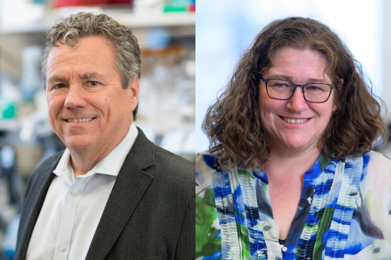
“It’s increasingly clear that the progression of a malignant tumor isn’t just about transformations happening within cells, but about coordinated interactions between cells and nearby tissues that develop ecosystems to support tumors,” says study co-senior author Scott Lowe, PhD, Chair of the Cancer Biology and Genetics Program, the Geoffrey Beene Chair for Cancer Biology at MSK’s Sloan Kettering Institute, and a Howard Hughes Medical Institute (HHMI) Investigator.
The work is representative of a broader “ecosystems” initiative at MSK, supported by the Marie-Josée and Henry R. Kravis Cancer Ecosystems Project, for which Dr. Lowe serves as scientific director. The initiative is focused on understanding cancer not simply as a genetic disease, but as an interconnected ecosystem of cells, tissues, and signaling networks. By framing cancer in this way, the program aims to enable the development of next-generation therapies that target not only tumor cells, but also the surrounding niche that supports their growth and progression.
Pancreatic Cancer: The Road to Malignancy
Pancreatic ductal adenocarcinoma is one of the deadliest cancers, with a five-year survival rate hovering around 13%. It also tends to develop through recognizable stages — from early benign growths to invasive cancer — making it a useful model for studying how cells transition from pre-cancer to cancer.
In pancreatic cancer, one genetic mutation stands out. The KRAS gene is altered in nearly all pancreatic ductal adenocarcinomas, the disease’s most common and deadly form.
When cells in the pancreas are injured by inflammation caused by, for example, pancreatitis, they normally enter a temporary repair program and then return to normal. But when the cells contain an oncogenic KRAS mutation, that repair program can get stuck in the “on” position, keeping cells in an unusually flexible state, instead of resolving normally.
Still, these precancerous cells with KRAS mutations can remain benign for a long time.
To find out exactly what is required for them to become malignant, the research team used a variety of advanced technologies at MSK:
- Genetically engineered mouse models.
- The latest molecular profiling tools — including single-cell RNA sequencing and spatial transcriptomics.
- Sophisticated computation to examine gene activity cell by cell, including changes to surrounding tissue.
A Tug-of-War Inside Cells: Oncogenes vs. Tumor Suppressors
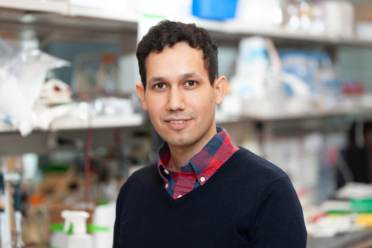
“Essentially, what we saw was a tug-of-war between oncogenes and tumor suppressor genes,” says the study’s lead author José Reyes, PhD, a postdoctoral researcher jointly mentored by Dr. Lowe and computational biologist Dana Pe’er, PhD, the study’s other senior author.
The team tracked pancreatic cells with KRAS mutations and used a marker that let them spot cells that had also lost the tumor suppressor p53. The p53 gene is known as the “guardian of the genome” because it turns on protective programs when cells show signs of DNA damage. Therefore, it was important for researchers to compare tumor cells to precancerous cells both with and without working p53.
Using these techniques, the scientists were able to identify a subset of precancerous cells that most closely resembled tumor cells — cells right at the tipping point of becoming malignant.
Counterintuitively, they found that these cells also had the most strongly activated tumor-suppressors: p53 and two others — CDKN2A and SMAD4.
This combination of opposing forces — a high oncogenic drive plus a high tumor-suppressor response — often results in a stalemate, putting cells into what scientists call “senescence,” a kind of emergency brake that prevents cells from continuing to divide when growth signals are abnormally strong.
“The body is seeing this particular cell state as dangerous — and putting extra pressure on it to stop,” Dr. Reyes says.
But if the body can’t get cells unstuck from this state, the cells begin to reshape their surroundings and set the stage for cancer to emerge — especially when additional genetic changes knock out p53.
“What we’re finding is that p53 is even more than the guardian of the genome, it’s the guardian of plasticity,” says Dr. Pe’er, Chair of MSK’s Computational and Systems Biology Program and an HHMI Investigator. “Cells need to become plastic for regeneration and repair to happen, but p53 is there to pump the brakes if they try to go rogue. Without p53, this flexibility can be hijacked by cancer.”
Dr. Pe’er also serves on the scientific advisory committee for the Ecosystems Project, and leads the computational and technology infrastructure for it.
Before a Tumor Forms, the Neighborhood Starts To Change
Pancreatic tumors are among the most difficult to treat because they’re protected by a dense weave of scarlike tissue and support cells.
The study shows that pre-cancers start to make these protective neighborhoods surprisingly early — well before a full-blown tumor appears.
The team was able to see how precancerous lesions and their surroundings change together using two techniques. The first is spatial transcriptomics, which allows researchers to visualize where specific cells and genes are active within a tissue. They also developed a new computational approach to modeling changes in the local neighborhood around a given cell.
“For the first time, this new approach allowed us to reconstruct dynamic changes in a cellular neighborhood — and allowed us to see how this rare subset of cells makes changes in nearby cells that support the development of a tumor,” Dr. Reyes says.
As researchers further investigated the cellular neighborhood, they discovered cells stuck in the injury repair state increasingly engage two key players, which undergo coordinated changes:
- Fibroblasts, which begin to build up fibrous, scarlike tissue.
- And myeloid immune cells, which start sending “stand down” signals that keep other immune cells from attacking.
“The neighborhoods around these pre-cancers start to look very much like the environment you see in invasive pancreatic cancer,” Dr. Pe’er says. “This repair program allows the pre-cancer to communicate with its neighbors to reshape its local environment in ways that pave the way for cancer to take hold.”
The new study builds on a previous collaboration between the Lowe and Pe’er labs that showed how cell-to-cell communication expands during the early development of pancreatic cancer.
A Critical Window for Intervention
One of the reasons pancreatic cancer has such a low survival rate is that it’s usually caught late, when the tumors are well-established and have started to spread.
The encouraging news is that this early ecosystem might present an opportunity for medical intervention, the researchers say.
The scientists gave mice a short treatment with a KRAS-blocking drug, which not only eliminated the premalignant cells, it disrupted the niche around them, too.
“A three-day treatment in mice paused cancer development for months, which would be the equivalent of years on a human timescale,” Dr. Lowe says.
For Dr. Lowe, who has studied p53 since graduate school, the work is a payoff for decades of research.
“For the first time, we’ve caught p53 in the act of suppressing cancer,” he says.
Not only do p53 mutations happen in about half of all cancers, those cancers are also among the most aggressive and often there are no good treatment options.
“So, it’s been a major goal of the field to develop strategies against these cancers,” Dr. Lowe says.
Many cancer drugs work by shutting down oncogenes that are overactive, but trying to compensate for p53’s loss has presented a much tougher clinical challenge.
“Some efforts have tried to mimic p53’s action or identify other vulnerabilities that make these cells cancerous,” he says. “These findings suggest that if p53’s job is to eliminate this plastic, injury repair state, then strategies to directly target this state and its protective niche could represent an innovative way to help the significant percentage of patients whose cancers have lost p53.”
Complementary Study Highlights Clinical Opportunities
Another recent study from Dr. Lowe, Dr. Pe’er, and their collaborators provided proof-of-principle evidence that supports the idea of targeting this injury repair state and the tumor niche.
Those findings, published March 30 in Cell, showed that these highly plastic cancer cells — which p53 normally eliminates and that expand following p53 mutation — express a cell surface molecule called uPAR. The study found, excitingly, that CAR T cells engineered to target uPAR can effectively eliminate this state, even when p53 is disabled.
The March study was overseen by Dr. Lowe; Michel Sadelain, MD, PhD, who recently moved his lab from MSK to Columbia University; and Aveline Filliol, PhD, a senior scientist in the Lowe Lab.
“This study highlights how understanding fundamental principles of cancer biology can reveal new therapeutic strategies, and we are excited to bring such approaches into the clinic,” Dr. Lowe says.
Additional Authors, Funding, and Disclosures
Additional authors on the paper include Isabella Del Priore, Andrea Chaikovsky, Nikhita Pasnuri, Ahmed Elhossiny, Jin Park, Philipp Weiler, Tobias Krause, Andrew Moorman, Catherine Snopkowski, Meril Takizawa, Cassandra Burdziak, Nalin Ratnayeke, Ignas Masilionis, Yu-Jui Ho, Ronan Chaligné, Paul Romesser, Aveline Filliol, Tal Nawy, John Morris IV, Zhen Zhao, Marina Pasca Di Magliano, and Direna Alonso-Curbelo.
The work was supported by numerous funding agencies, including multiple grants from the National Institutes of Health and National Cancer Institute (5R01-CA283378-02, F31 CA290899, K00 CA245471, K08-CA255574, P30CA045508, R01-CA260752, R01-CA264843, R01-CA268426, R01-CA271510, U01-CA224145, U01-CA274154, U54-CA274371, U54-CA274492). Additional support was provided by the Howard Hughes Medical Institute, The Marie-Josée and Henry R. Kravis Cancer Ecosystems Project, Damon Runyon Cancer Research Foundation, Mark Foundation, Pancreatic Cancer Action Network, Cycle for Survival, MSK Center for Experimental Immuno-Oncology Scholars Program, and a Marie-Josée Kravis Fellowship in Quantitative Biology. For a full list of funders, please refer to the journal article.
Unrelated to the study, Dr. Lowe declares competing interests outside consultancy and equity for Oric Pharmaceuticals, Blueprint Medicines, Mirimus, Senecea Therapeutics, Faeth Therapeutics, and PMV Pharmaceuticals; and outside consultancy without equity for Fate Therapeutics. Dr. Pe’er reports equity interests and provision of services for Insitro, Inc. Dr. Romessor provides compensated professional services and activities for EMD Serono, Faeth Therapeutics, Urogen Pharma, Incyte, and Natera Inc; he also offers uncompensated professional services and activities for 10x Genomics, XRad Therapeutics, and the HPV Alliance and Anal Cancer Foundation nonprofit organizations. Dr. Chaligné is on the Scientific Advisory Board of Sanavia Oncology and LevitasBio and a compensated consultant for the Gerson Lehrman Group.
Read the study: “Oncogenic and tumor-suppressive forces converge on a progenitor niche at the benign-to-malignant transition,” Cell. DOI: 10.1016/j.cell.2026.03.032