As part of MSK’s 38th annual academic convocation on May 18, the Gerstner Sloan Kettering Graduate School of Biomedical Sciences (GSK) will award PhD degrees during its sixth commencement ceremony to Emily Casey, Marta Kovatcheva, Shefali Krishna, Daniel Marks, Anna Sophia McKenney, Prashant Monian, and Alison Spencer. Their graduation brings the total number of GSK alumni to 44.
Throughout the past year, the students successfully defended their dissertations — the final requirement in earning their doctorates. The promising work of this year’s degree recipients embodies GSK’s mission to train its students to tackle real-life clinical challenges through biomedical investigation. Learn more about their research below.
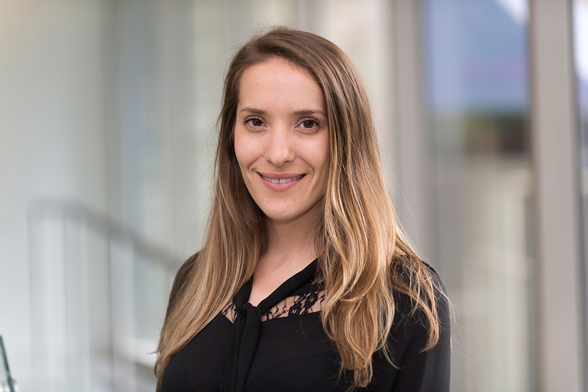
Emily Casey

Emily completed her thesis work in the laboratory of David Scheinberg. She generated a new mouse model to better predict the efficacy of human anticancer antibodies.
Clinical evidence suggests that the activity of most monoclonal antibody (mAb) therapies relies on interactions with Fcγ receptors (FcγRs) expressed by immune cells. Current preclinical models of mAb therapy for human cancers studied in mouse models utilize the mouse immune system for FcγR recognition and subsequent therapy. However, there are key differences between the mouse and human FcγR networks that do not allow for the complete and accurate understanding of mAb activity.
To address this issue, Emily generated an immunodeficient mouse strain in which human cancers can be studied. The mouse strain expresses the human FcγR repertoire instead of the mouse FcγRs. In an exciting result, this mouse model was able to accurately replicate human clinical data for comparing two mAbs for the treatment of human lymphoma, whereas a commercially available mouse model expressing the mouse FcγRs did not.
She also developed additional mouse strains that demonstrated great use in parsing out the mAb mechanisms of action in a model of thrombocytopenia, a low blood platelet count caused by mAbs.
Overall, these immunodeficient mice strains expressing human FcγR allow for the accurate and in-depth study of therapeutic antibody activity and the mechanisms of action against human cancers.
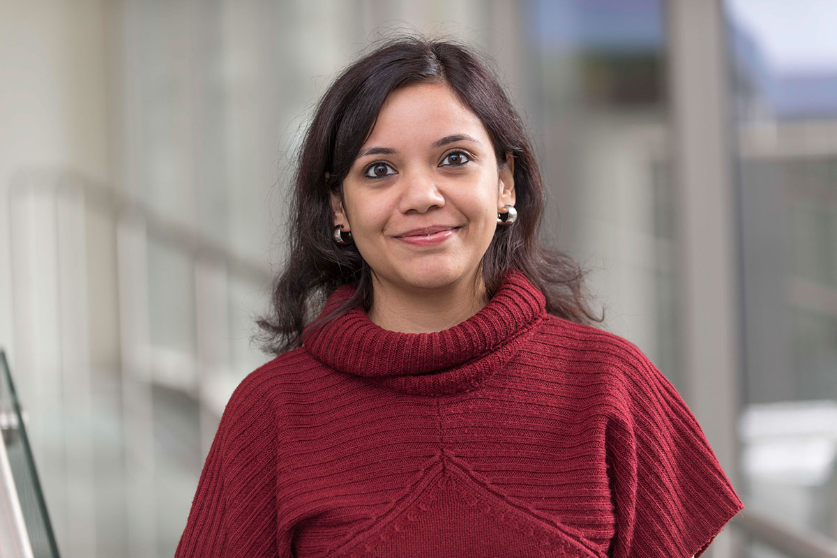
Marta Kovatcheva
Marta completed her dissertation in the laboratory of Andrew Koff, exploring the molecular mechanism of a new class of small molecule cancer drugs called CDK4 inhibitors (CDK4i).
CDK4 is a master regulator that drives the cell cycle forward and enables cells to divide. The majority of human cancers have in some way overactivated CDK4 signaling to promote their unrestrained division.
Despite the marked success of CDK4i in clinical trials, patients have not responded uniformly nor as preclinical models have predicted. By studying CDK4i in a rare cancer type known as well-differentiated and dedifferentiated liposarcoma (WD/DDLS), Marta uncovered that CDK4i could induce two distinct forms of growth arrest: quiescence, a reversible form of cell cycle arrest, and senescence, a permanent form of cell cycle arrest.
Marta observed that cells must reduce the level of a protein called MDM2 to undergo senescence in response to CDK4i. She tested the clinical relevance of these results using patient samples from a clinical trial in people with WD/DDLS at MSK and found that reductions in MDM2 in post-treatment biopsies correlated with a favorable clinical outcome. Thus, MDM2 serves as a biomarker for CDK4i efficacy.
Marta also discovered a novel role for an enzyme called ATRX in regulating senescence. She found that ATRX represses another growth-promoting gene called HRAS in response to CDK4i. HRAS activity can be targeted through already approved cancer drugs, suggesting combination therapies to be tested in the future.
Shefali Krishna
Shefali completed her graduate thesis in the lab of Michael Overholtzer. She focused her research on the regulation of lysosome function, which is required for the recovery of nutrients to support cell growth.
The rapid proliferation rates of solid tumors often outpace the supply of blood vessels, causing a metabolic crisis in tumor cells and an increased dependence on alternative pathways to gain nutrients. Cancer cells can engulf other cells and proteins to support their survival during starvation conditions. Once ingested, the extracellular cargo is degraded by the lysosomes and the nutrients are exported.
Shefali’s work builds on her previous finding that kinase mTORC1 regulates the size of lysosomal compartments in a cell and redistributes the digested cargo in the lysosomes. This occurs through a process called membrane fission, by which small vesicles bud off from the large lysosomal compartment, thereby shrinking its size.
Her thesis research identified another kinase called PIKfyve as a regulator of fission and cargo redistribution. She demonstrated that PIKfyve is required for the export of certain nutrients from the lysosomes, which could play a role in maintaining lysosome size and morphology and supporting the survival and proliferation of Ras-mutant cancer cells during starvation.
Her findings point to PIKfyve as a critical regulator of lysosome trafficking and nutrient homeostasis in cells. Her work also sheds light on the regulation of an alternative pathway of nutrient acquisition by tumor cells through the kinase PIKfyve. These results have important implications for the treatment of metabolically stressed tumors with Ras mutations, like pancreatic cancer, and recognize PIKfyve as a novel therapeutic target for these malignancies. Given that the PIKfyve inhibitor apilimod is currently in clinical trials for other diseases, this drug may have potential therapeutic value for patients with Ras-driven cancers.
After graduation, Shefali will pursue postdoctoral research at the Salk Institute, where she plans to investigate the role of nuclear membrane proteins in cancer and aging.
Daniel Marks
Daniel completed his graduate research in the laboratory of Robert Benezra, where he studied mitotic checkpoints, the control mechanisms that ensure proper division of a cell.
One of the hallmarks of cancer is its ability to change and adapt very quickly. Cancer can mutate through faulty separation of replicated DNA during mitosis, or cell division, which can lead to large gains and losses of genetic information. Previous research conducted in Dr. Benezra’s lab has suggested that this abnormal cell division process may be due to dysfunction in the mitotic checkpoint caused by overexpression of checkpoint genes.
Daniel studied how TRIP13 — a newly identified regulator of the mitotic checkpoint — plays a critical role in this process. He found that inhibiting TRIP13 in cells that overexpress these checkpoint genes could specifically block the exit from the division phase of the cell cycle.
His findings point to TRIP13 inhibition as a potential therapeutic target in cancer cells that have a dysfunctional mitotic checkpoint, which possibly offers a more precise way to disrupt cell division.
Anna Sophia McKenney
Sophie is the first student enrolled in the Tri-Institutional MD-PhD Program to receive her PhD from GSK. She completed her dissertation research in the laboratory of Ross Levine, using mouse and human models to focus on the cause, development, and treatment of a group of benign blood disorders called myeloproliferative neoplasms (MPN). She set out to learn more about what happens when MPN progresses to acute myeloid leukemia (AML), an often deadly type of blood cancer.
While MPNs are usually characterized by a mutation in the protein JAK2, Sophie observed a new mutation in the protein IDH among individuals whose disease developed into AML. She created a mouse model that combined these two mutations and used drugs that specifically targeted the action of both JAK2 and IDH to make the disease regress in both mouse models and patient samples.
She showed that this combination of mutations in mice can lead to cancer and that drugs targeting these mutations have the potential to reverse the disease. A clinical trial is currently being initiated to test this novel regimen in people with MPN who harbor the two mutations.
After defending her thesis, Sophie returned to medical school to earn her medical degree and apply for her residency.
Prashant Monian

Working in the lab of cell biologist Xuejun Jiang, Prashant focused his dissertation research on the mechanisms underlying different forms of cell death, which is essential to maintain homeostasis within an organism. Aberrations in the cell death process can lead to various diseases, including cancer. Prashant’s thesis examined the regulation and mechanism of two distinct cell death processes: apoptosis and ferroptosis.
Apoptosis is dependent on the activation of caspases, a family of enzymes that trigger the normal breakdown of proteins. Prashant identified a novel signaling pathway that amplifies the apoptotic signal by increasing the expression of the cellular apoptosis susceptibility protein (CAS), which in turn leads to increased caspase activity. He also found that CAS is overexpressed in a subset of human breast tumors, which could make tumor cells more sensitive to certain apoptotic attacks. These findings suggest that CAS plays contrasting roles in proliferation and apoptosis and that overexpression of CAS in tumors could serve as a potential biomarker to guide therapeutic choices.
In the second part of his dissertation, Prashant explored the mechanism of ferroptosis, a recently identified form of iron-dependent cell death. He found that ferroptosis is accompanied by biological changes in the architecture of the cell’s mitochondria, the specialized structures responsible for creating the energy needed to sustain life and support organ function. He demonstrated that the presence of mitochondria with an intact membrane contributes to ferroptosis. These findings implicate a central role for mitochondria in ferroptosis and could lead to the identification of new therapeutic targets.
Alison Spencer

Alison completed her dissertation work in the lab of Jennifer Zallen. Using Drosophila embryos, she set out to better understand how cells build and organize tiny subcellular structures in epithelial cells that are involved in larval movement.
Denticle precursors are micron-scale, spike-shaped projections that are distributed in rows across the lower surface of the fruit fly larva. They are rich in actin, a protein present in all cells and in muscle tissue, where it plays a role in contraction. Alison studied how the organizational patterns of these structures are created and maintained during the dramatic changes in cell size that occur during larval growth.
Alison used quantitative imaging techniques to shed light on the mechanisms by which actin patterns are generated inside a cell. She also developed computational methods to quantify the spatial patterns in which denticle precursors are organized. Additionally, she identified and characterized several mutants that alter or fail to establish this precise cytoskeletal pattern.
Her findings demonstrate that denticle spacing is not random. Denticles are precisely positioned within cells and this organization scales with cell size. The number and the spacing of denticle precursors within cells are reduced under space-limited conditions and robustly increase during larval growth, and the accuracy of denticle organization requires an intact microtubule network. These results define a novel mechanism for microtubule-dependent actin scaling that maintains precise spatial patterns of actin organization during tissue growth.