Inspired by a surprise discovery, Memorial Sloan Kettering scientists have engineered a tiny particle that could make it possible to deliver drugs directly to the kidneys and minimize their uptake in other organs. The device, called a mesoscale nanoparticle, could help boost the usefulness of some kidney cancer drugs and might also be used in the treatment and diagnosis of other kidney conditions.
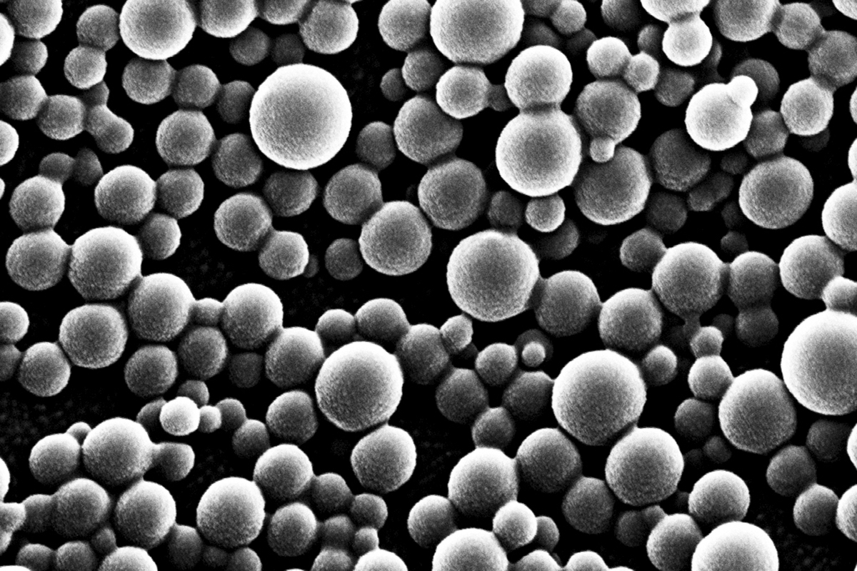
Nanoparticles are polymers — large molecules consisting of many small parts — that typically measure between one and 100 nanometers and can be loaded with drugs or imaging agents. (For comparison, an average human cell measures about 50,000 nanometers in diameter.) They hold promise for many medical applications, partly because they are biocompatible — not harmful to living tissue — and have unusual chemical and physical properties.
At MSK and elsewhere, scientists are seeking to develop new nanoparticles capable of ferrying drugs to tumors and making them stay there long enough to be effective. “Targeting tumors specifically has often proved to be challenging,” says chemist and engineer Daniel Heller, who led the study. “For cancers growing in specific sites, such as the kidneys, the next best thing may be a particle that’s capable of seeking out the right organ.”
A Serendipitous Result
The kidney-bound particle is the result of an incidental finding made while studying how the size and chemical properties of some nanomaterials could be modulated to guide their distribution in the body. The goal of the project — a collaboration between Dr. Heller’s group and MSK lung cancer physicians — was to create a nanoparticle capable of targeting cancer drugs to the lung.
The investigators produced a number of nanoparticles of different sizes and chemical properties. They then injected these particles into mice and tracked their location using CT scans and other imaging methods.
The results of the initial experiments were mixed. In particular, one of the particles didn’t behave as expected. The researchers could find only low levels of it in the animals’ lungs, and very little in the liver or spleen, where many similar substances tend to gather. “To our surprise, this particle accumulated almost exclusively in a specific structure of the kidney,” says postdoctoral fellow Ryan Williams, the study’s first author, “and it stayed there until all of it had degraded.”
The researchers dubbed the particle “mesoscale” or middle-size because it’s bigger than most of its forerunners, with a diameter of about 400 nanometers. It takes up to two months for the mesoscale nanoparticle to fall apart completely, which means it could potentially be used to release a drug very slowly inside the kidneys.
Redeeming Failed Drugs
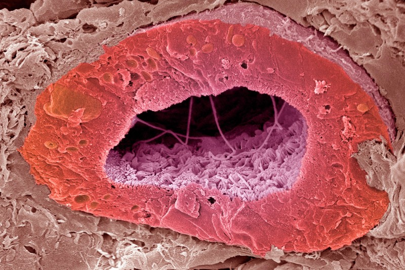
Located right below the rib cage, the kidneys are two fist-size organs whose main function is to filter waste and excess water out of the blood to make urine. While waste products enter the kidneys through a network of filtering blood vessels called the glomerulus, the researchers found that mesoscale nanoparticles are absorbed — through a poorly understood process — in a different part of the kidney called the proximal tubule.
“This is interesting for several reasons,” Dr. Heller says. “First, the proximal tubule happens to be where renal cell carcinoma [RCC, the most common form of kidney cancer] originates.”
The team is now working to develop a nanoparticle-based technology to deliver chemotherapy or targeted therapies directly to the site of RCC. They hope the method will help reduce side effects of the drugs by keeping them away from other organs.
RCC drugs that haven’t met expectations could potentially be improved with nanotechnology.
“We are currently doing experiments to verify that the particles can be used to effectively deliver a drug to RCC tumors in mice,” Dr. Heller says. The main focus of this work, he adds, will be on kidney cancer drugs that have failed clinical trials or preclinical testing.
“The drugs that have been shown to work already are presumably hitting their target,” he explains. “So it might make more sense to focus on compounds whose development has been halted, either because they didn’t work as well as people had hoped or because they caused too many side effects. There’s a chance we could rescue the promise of some of these drugs by delivering them directly to the tumor site.”
More Possible Applications
The researchers are exploring other potential uses of the particle as well. For example, they believe it could be employed to help repair kidney failure, a common problem in people who receive chemotherapy for various types of cancer. “In these patients, the proximal tubule is often the first site of damage,” Dr. Heller says.
In collaboration with nephrologist Edgar Jaimes, who heads MSK’s Renal Service, he and Dr. Williams are investigating whether this technology could potentially be used to develop therapies to accelerate kidney recovery or prevent chemotherapy-induced kidney injury. They will soon start experiments to find out if the particles can deliver small molecules that target proteins involved in acute kidney injury. They will first explore the approach in animal models with the ultimate goal to determine its usefulness in patients with chemotherapy-induced kidney injury.


