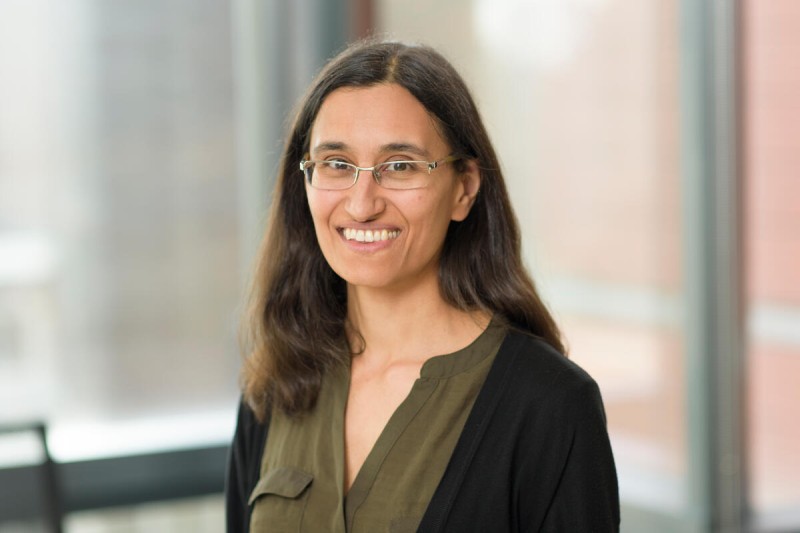
Computational biologist Christina Leslie, PhD, has spent nearly two decades at Memorial Sloan Kettering Cancer Center (MSK) building computational tools that help make sense of some of the most complex data in biology.
Today, Dr. Leslie’s work sits at the crossroads of two technological revolutions. Single-cell genomics is allowing scientists probe the activation of genes in individual cells revealing the distinct roles, identities, and behaviors of every cell type in a tissue or tumor. Meanwhile, the rise of artificial intelligence (AI) is making it possible to find meaningful patterns in hugely complex single-cell datasets at a scale far beyond more traditional computational methods.
We sat down with Dr. Leslie to discuss the promise — and limitations — of this frontier, and how these advances could help researchers design better therapies, decode why tumors resist treatment, and tailor treatments to each individual and their unique cancer.
Your lab works at the intersection of AI and biology. How would you describe what you study?
The theme of my lab, which is in MSK’s Sloan Kettering Institute, is studying how gene expression is regulated in mammalian cells — including in the immune system and in cancer. We use tools from what used to be called machine learning and is now called artificial intelligence.
In the past few years, we’ve been using AI to analyze three increasingly powerful types of biological data.
- The first is single-cell data: Rather than working with millions of cells all mixed together, we can now read the genetic activity and epigenetic state of individual cells one at a time, revealing what each cell type is actually doing within a tissue or tumor.
- The second is spatial transcriptomics, which takes that one step further. It tells us not just what genes are active in individual cells, but where those cells are physically located in a tissue sample.
- And the third is 3D genomics, which maps how DNA is physically folded and organized inside the cell nucleus. This is important because the physical shape of DNA influences which genes get switched on or off.
AI gives us the computational power to make sense of all three of these extraordinarily complex data types and to find the patterns within them that would otherwise be invisible.
Single-cell genomics has been described as a revolution in biology. Why does it matter so much?
Single-cell genomics is an increasingly important way that we make new discoveries in biology. Before, we would do what’s known as “bulk” analysis, which allowed us to look at what was happening in a tissue sample — but with the molecular data from all the cells mixed together. Now, we have ways of barcoding individual cells in a sample so that we can understand how cells play different functional roles in a particular system. And we can discover new cell types and functional specializations.
Let me give you two examples from recent work done at MSK:
We collaborated with Dr. Chrysothemis Brown using single-cell approaches to discover a new specialized type of immune cell called Thetis cells, which help explain how the immune system learns to tolerate food and helpful gut bacteria early in life. This work is shedding new light on childhood food allergies.
We also worked with the lab of Dr. Alexander Rudensky, who heads MSK’s Immunology Program, to uncover that there are two distinct types of regulatory T cells at work in colorectal cancer — IL-10 positive and IL-10 negative — which play completely opposite roles in tumor progression. One promotes tumor growth while the other restrains it — which highlights the need to develop therapies that can selectively eliminate the harmful cells while preserving the helpful ones. We wouldn’t have been able to differentiate between these two subpopulations of cells without single-cell techniques.
How does AI connect to all of this?
Datasets generated by these technologies are quite large — some single-cell atlases are now 150 million cells — you need more sophisticated tools to make sense of them.
The AI community adapted models from natural language processing and computer vision for single-cell data relatively early on. And there’s real logic to that — you have a lot of data, you need to find patterns, and these tools are powerful.
But cells, genomes, and expression profiles are not natural language and they’re not images. If you just directly take these models and say, “OK, we have a bunch of genes, so let’s treat them like words” — that’s not quite right. Genes aren’t organized in a sentence. There is an underlying structure, which is the structure of gene regulation. When cells receive specific signals from the body, they turn on sets of genes in a coordinated way. And that biology needs to be represented in our models.
How would you describe the state of the field right now?
The current models have been made by serious people doing serious work — they know their AI. But they’re just too generic as representations of single-cell data, and they’re not solving tasks that biologists conducting experiments really need. It would be amazing if you could train a model on 150 million cells and then automatically, correctly label all the cell types in your new dataset. That’s not possible yet. The level of automated annotation from these models is too coarse-grained — that is, too broad and imprecise.
To move the field forward, I think there needs to be more communication between bench scientists and computational researchers about what the real problems are — and then we can work to solve them together. I recently co-authored a review article on this exact topic with two of my mentees, postdoctoral researcher Sneha Mitra and graduate student Jiaxin Li.
What about virtual cells — the idea of simulating biology in a computer? There’s been a lot of excitement there.
The virtual cell is perhaps partly a branding exercise — I don’t know if we’re allowed to say that — but the core idea is genuinely compelling. What if you could use AI to predict how a cell will respond to a drug or to a genetic change before running a single experiment in the lab?
In biology, we call those interventions “perturbations” — anything that disrupts or changes a cell’s normal state. That could be a drug, a cytokine used by the body to signal the immune system and fight infection, or deliberately switching off a particular gene. Scientists have generated enormous datasets doing exactly this — exposing cells to different drugs or genetic changes and measuring what happens — and the hope is that an AI model trained on all of that data could learn the rules well enough to make accurate predictions about interventions it has never seen before.
Imagine being able to ask: If I give this cancer cell this particular drug, what happens? — and getting a reliable answer from a computer model rather than spending months in the lab. That would be transformative.
We’re not there yet. These models are improving, but they don’t yet account for enough of the real biology to be reliably predictive. With better modeling that includes a more robust understanding of how genes are regulated, I’m hopeful we can get there.
What would it take?
What we study is how genes are regulated — how the language in non-coding elements of the genome allows transcription factors to bind and orchestrate gene expression. A piece of what’s missing in current virtual cell models is understanding how perturbations relate to each other. Perturbations affect different pathways, and we have some information about how those pathways connect to downstream transcriptional responses — in other words, the cascade of changes in gene activity that follow. Encoding some of that should help.
Right now, a lot of models just say: Encode the perturbation, encode the cell, add the representations together, and that’s your prediction. It’s too simplistic. The models need to better represent the biology of how cells make decisions — not just compress the data into an AI algorithm and hope the correct answer comes out the other end.
You spoke of a gap between the AI research community and the experimental biology community. How do you bridge that?
It really is a two-way street. Our collaborators teach us about the latest problems in immunology or cancer biology, and that informs how we model. But then we can sometimes find answers in the data that help drive a project forward. The key is communication — and being willing to define success by what biologists actually need, not just by whether a model sets a new benchmark on a test that the AI community designed for itself.
At MSK, I tell students who are interviewing for our PhD program: You won’t come here and work on the wrong problem. You won’t solve something that’s computationally interesting but doesn’t actually matter — because you’re in constant communication with top immunologists, top cancer biologists. You see the latest of what’s happening in the lab, and you think more critically about what the computational side of things should contribute.
Can you give an example of what that close collaboration looks like in practice?
Sure. We had a project studying B cell biology — germinal center B cells — using single-cell multiome data generously shared by the lab of Dr. Ari Melnick at Weill Cornell Medicine. We made the call early on that we were going to tell a biologically interesting story, not just write a methods paper.
The research centered on a gene called Arid1a, which plays an important role in regulating how DNA is organized and read inside a cell. When Arid1a is lost or damaged — which happens in certain B cell lymphomas — it disrupts the normal process of B cell maturation, pushing cells toward an abnormal state associated with cancer. Understanding how to reverse that process could point toward new treatment strategies.
Using our AI tool, DynaVelo, we were able to run virtual experiments — essentially asking the model: If Arid1a is lost, what genetic change might restore normal cell behavior? The model nominated three candidates. Then our collaborator, Dr. Darko Barisic at Weill Cornell, generated the mouse models needed to test our predictions, ran the experiments, and showed that in each case the changes DynaVelo identified really did restore normal B cell behavior. All three predictions held up.
That’s the power of this approach — instead of spending years testing every possibility in animal models, we could screen thousands of candidates virtually and take only the most promising ones into the lab. And that’s what’s possible when the computational and experimental sides are genuinely working together.
What else are you excited about on the horizon?
One thing I’m really excited about is a new approach to spatial transcriptomics — examining the activity of genes inside a tissue, cell by cell, while keeping their precise physical location intact.
Dig Vijay Kumar Yarlagadda, a student in my lab who is co-advised by Dr. Joan Massagué, has a computer vision background and developed a completely new way of analyzing imaging-based spatial transcriptomics data. The model is called Nicheverse. We’re using it for a project on renal cell carcinoma metastasis and in another on how pancreatic cancer spreads along nerve cells.
And that’s typical of what’s possible at MSK — because of the collaborations, the quality of students, and embedded computational research in a biomedical environment.
Finally, for people who haven’t been following this field as closely as you have, what’s the biggest idea you want them to take away?
The promise of AI to transform biology is real, and it’s important — but it needs to be done right.
The data resources we have now are extraordinary. The question is whether we can build models that are smart enough, and biologically grounded enough, to help laboratory scientists and clinicians make discoveries. We’re not there yet for some of the most ambitious goals, but the path is becoming clearer.
Dr. Leslie is the Program Chair of the Tri-Institutional PhD Program in Computational Biology and Medicine.
Dr. Leslie holds the Virginia and Daniel K. Ludwig Chair at MSK.