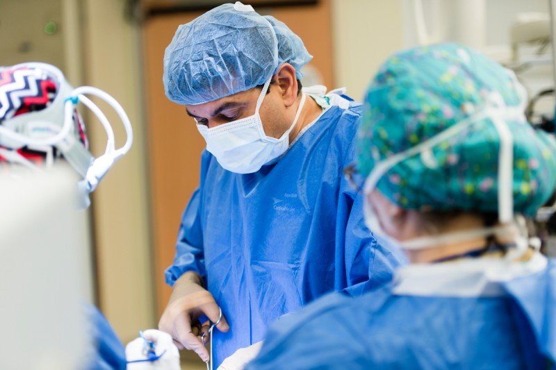
Surgery is the primary treatment for most head and neck cancers. These cancers are often near critical nerves and blood vessels that control important bodily functions, so removing them is especially challenging. In some cases, these cancers may actually invade organs, such as the tongue or voice box.
Fortunately, new imaging technologies are being investigated at Memorial Sloan Kettering. These methods will allow surgeons to see cancer more precisely in delicate tissue, especially in the case of mouth cancer and tongue cancer. Head and neck cancer surgeon Snehal Patel explains how innovative technology could help surgeons like him plan and perform operations to treat these cancers.
Why are imaging approaches especially important for surgery on mouth cancer and tongue cancer?
Diagnosing mouth cancer and tongue cancer at an early stage can be difficult. Precancer and early cancer often look similar to other common lesions. Tobacco and alcohol use, both major risk factors for these cancers, can complicate a diagnosis because they alter the appearance of tissues in the mouth. To get a clear diagnosis, surgeons usually have to do a biopsy. Then a pathologist will examine the sample under a microscope. Knowing where to take the biopsy can be challenging even for experienced doctors. It is often hard to tell what is cancer and what is normal in this part of the body.
Even after a diagnosis, the surgery itself can be complicated. To make sure that all of the cancer is removed, a surgeon takes an extra margin of normal-appearing tissue — up to a centimeter and a half — around the cancer. In the mouth or tongue, any extra tissue loss can impair the ability to speak or swallow. That can have a huge impact on a person’s quality of life. So it’s critical to find better ways for a surgeon to see exactly where the cancer is to be able to remove the entire tumor while sparing as much normal tissue as possible.
What new imaging approaches are being developed?
We are exploring three novel techniques in clinical trials. Two of them help delineate the presence and extent of cancer in the mouth and tongue. The third technology involves the use of nanoparticles to more precisely see cancer that may have spread to lymph nodes. All of these technologies were generated here at MSK or, in some cases, in collaboration with researchers and doctors at Weill Cornell Medical College.
Could any of these imaging techniques allow for earlier diagnosis of oral cancers?
I am collaborating with MSK radiochemist Thomas Reiner on developing and testing a compound called PARPi-FL. It can detect and illuminate cancer cells. PARPi-FL contains an imaging agent that naturally binds to a molecule called PARP, which is abundant in cancer cells. The agent is attached to a fluorescent molecule that glows when exposed to a laser. This signals the presence of cancer cells, which can be confirmed by a biopsy. We can use this compound as a mouthwash to mark the tumor edges and guide us during surgery. It also shows the presence of residual cancer cells in the surgical bed after the removal of the cancer.
The mouthwash can actually be produced as a powder that does not need refrigeration. It can be mixed on the spot at virtually any location. This technology has a huge potential for effective large-scale screening for early detection of oral cancer in dentists’ offices and other settings, including countries that lack many medical resources.
What other new approaches can make biopsies and surgeries more precise?
Another trial we are conducting tests a technology called reflectance confocal microscopy (RCM). This makes it possible to do a real-time optical biopsy in a person’s mouth without cutting tissue. RCM is a handheld, laser-based technology that allows us to identify cellular and structural details in tissue without removing it for examination in a pathology lab. We insert a rod-shaped device into a person’s mouth. The laser then scans the surface of the tissue and collects the reflected information. Using this probe, we are able to capture still and video images — of a lesion on a tongue, for example — in microscopic detail. We can electronically send those to a central facility for examination and diagnosis by expert pathologists. Anything that looks suspicious could be followed up with a conventional biopsy for confirmation of a cancer diagnosis.

In addition to possibly helping with the early detection of cancer, this will be especially useful during surgery. The standard method of analyzing tissue requires us to remove a sample. Analysis under a microscope takes up to four or five days to complete. With RCM, I can envision a situation in which I can transmit multiple images or videos of different sections to a pathologist in real time — during an operation — and we confer on where the cancerous tissue stops and normal tissue begins. That would allow me to be more precise when I’m removing the cancer to maximize the chances of cure and at the same time spare as much normal tissue as possible.
How is imaging being used to track cancer that has spread?
Head and neck cancers typically spread to the lymph nodes in the neck first. I am working with clinician-scientist Michelle Bradbury to test a new optical-imaging approach that allows us to more precisely see this type of cancer spread. This minimally invasive technique combines the use of nanoparticles known as C dots with a handheld camera that detects fluorescent light emitted by the nanoparticles when they are illuminated by a special laser. This creates a high-resolution image showing where the cancer cells are. The surgeon can then remove lymph nodes with minimal disturbance of adjoining nerves and blood vessels.
How soon will these investigational techniques become available to head and neck cancer surgeons?
All MSK patients with these cancers are offered the treatments if they meet the criteria for inclusion in the clinical trials. The PARPi-FL and RCM technologies are currently being tested for oral cancers. We envision using them in combination at some point in the future. The C dots lymph node study is being conducted in people with head and neck melanoma. We expect this technology to be useful for a number of head and neck cancers, including oral cancers. In short, we hope these will be implemented into standard MSK clinical care within a few years.
We are pleased that we can offer these innovative and cutting-edge cancer detection technologies as an option to our MSK patients right now. We are confident that these technologies will eventually change surgical practice around the world. Surgeons often resist using something that is brand new. If we are able to prove that these techniques work, it would have widespread benefits for screening, early detection, and image-guided minimally invasive surgical therapy.

