T cells are the trained killers of the immune system and the driving force behind cancer immunotherapy. But the behavior of T cells is hard to predict: Some spring into action when prompted, while others become lethargic and aimless. This puzzling inconsistency is one of the main reasons immunotherapies don’t work in most cancer patients.
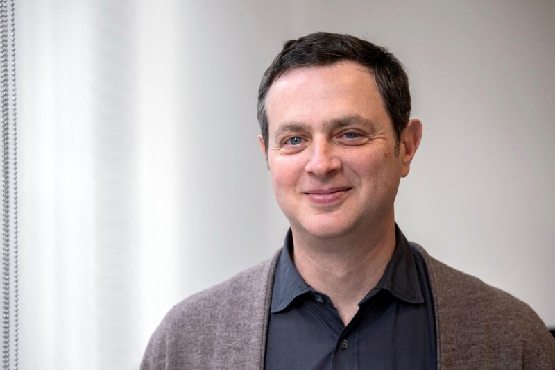
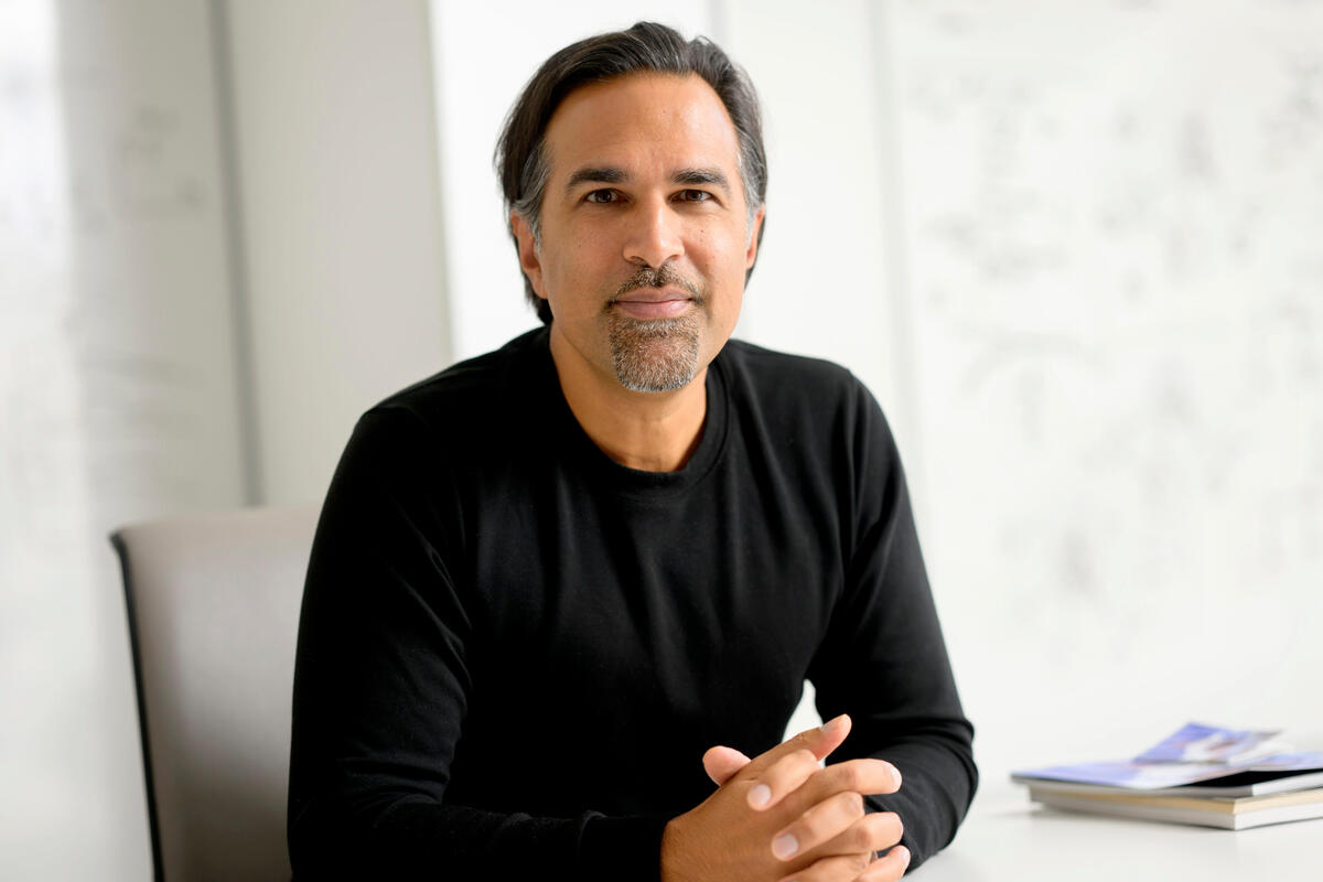
To tackle this problem, Memorial Sloan Kettering Cancer Center (MSK) researchers have developed a new approach to better understand and predict T cell behavior. The team, led by computational biologists Benjamin Greenbaum, PhD, and Sohrab Shah, PhD, designed a statistical model to monitor T cell responses to immunotherapies, including therapeutic cancer vaccines.
By connecting a T cell’s receptors — which can recognize antigens (protein fragments) on the surface of cancer cells — to how it behaves, the researchers hoped to address a fundamental question: How does a T cell’s genetic identity (the unique genetic sequence of its receptor) relate to its biological behavior — and how does that behavior change with immunotherapy?
The researchers used a new computational method, called TCRi (for TCR information), to analyze data from more than 167,000 T cells from cancer patients who had received immunotherapy drugs called checkpoint inhibitors.
“This approach produces a standardized measurement that lets us compare results across different studies and clinical trials,” Dr. Greenbaum says. “We have a way to learn from this data what a meaningful T cell response to an immunotherapy looks like — or doesn’t.”
The researchers report their findings in Nature Genetics. The co-first authors were Nicholas Ceglia, PhD, and Sohrab Salehi, PhD, in the laboratory of Dr. Shah, and Zachary Sethna, PhD, in the Olayan Center for Cancer Vaccines at MSK, directed by Vinod Balachandran, MD.
T cell receptors as highly specific locks
T cell receptors can recognize and bind to certain antigens present on the surface of cancer cells. Like a key turning a highly specific lock, the interaction between antigen and receptor will trigger a T cell to make copies of itself with identical receptors that can recognize the same antigens. This trait is known as receptor clonality.
The presence of many T cells sharing the same receptor suggests a strong anti-cancer immune response. But the existence of this newly created T cell army can be misleading: Even when the T cells seemingly have the right receptors, they often fail to act when prompted by immunotherapy.
“Looking at clonality alone can lead you to confuse responders with non-responders,” Dr. Greenbaum says. “The T cells of non-responders can look very clonal, but they don’t behave as if they can still recognize the antigens they should. This new tool can help give us a clearer understanding of T cell behavior.”
Focusing on the relationship between receptors and T cell state
The new model designed by the researchers could shed light on T cell behavior by examining the relationship between the makeup of these receptors and the actual state of the T cell.
The research draws on information theory, a branch of mathematics created by Claude Shannon in 1948 that studies how information is measured, stored, and sent. (Anthropic named its artificial intelligence tool “Claude” in his honor.) Information theory treats information itself as a measurable, statistical quantity like mass or energy.
The MSK researchers were able to measure the information using single-cell technology, which allows them to study individual cells and see which genes are active. The technique analyzes the relationship between the distributions of two different features of the same T cell:
-
One feature is the genetic sequence of the T cell receptor, which is like a unique genetic barcode that determines what the T cell can recognize.
- Another feature is the cell’s gene expression pattern. It reveals what kind of state the cell is in and what it may be doing. For example: Is the T cell active, primed to recognize a cancer cell, exhausted (dysfunctional), or completely “naive” — meaning it hasn’t yet encountered its specific target antigen?
TCRi essentially measures the strength of the correlation between a T cell receptor’s “clonotype” (the barcode) and its gene expression pattern (the cell state or “phenotype”). In other words, if you know a receptor’s identity, how well can you predict a T cell’s state?
“Our approach can help us understand T cells more completely by combining two kinds of information from a single cell within the mathematical framework of information theory,” Dr. Greenbaum says. “Essentially, we are looking at how well the T cell’s receptor is communicating with its cell state. At a large scale, we can quantify data about T cell response — or non-response — to therapies that we are testing in clinical trials and immune monitoring efforts.”
TCRi offers a more rigorous analysis than previous methods
The advance represented by TCRi is that it combines the two pieces of information in a balanced way within the framework of information theory. Instead of saying a given T cell is “type X,” it can assign probabilities, such as saying a cell is 80% likely to be one state and 20% another. This gives a more accurate overall picture for large amounts of data.
In analyzing the data from more than 167,000 T cells from cancer patients who had received checkpoint inhibitors, the researchers wanted to see if there was a difference between responders and non-responders (patients in whom T cells were not activated).
One result was a bit unexpected. They found that a lower correlation between receptor and cell state made the T cells more likely to respond to immunotherapy drugs.
-
In non-responders, the T cells’ clonotype and phenotype were more tightly correlated and less open to outside influence. This tight connection existed before treatment started, and the link often increased after therapy began.
- In responders, the link between clonotype and phenotype was looser prior to treatment — the T cells were in a more “receptive” or flexible state that allowed more productive responses when the immunotherapy unlocked them.
This finding sheds more light on T cell behavior, including which types of T cells might be more likely to respond to immunotherapy drugs.
Dr. Greenbaum, who is also Co-Director of Neoantigen Discovery at the Olayan Center for Cancer Vaccines at MSK, says TCRi has already been used at MSK to monitor a patient’s response to vaccines, including an investigational personalized pancreatic cancer vaccine being tested at MSK.
In a phase 1 trial, 50% of vaccinated patients responded to the vaccine. Six out of eight who responded remained cancer free after six years of follow-up. Understanding why some patients respond while others don’t is important to extending the potential benefit of a promising treatment.
“This tool offers a way to get a much clearer picture of what is biologically meaningful in T cell behavior,” Dr. Greenbaum says. “Not only is it helping us to better understand and predict the initial response to cancer vaccines, we also are monitoring how the T cell receptors — and the cell states — change over time as a result of the vaccine. Being able to monitor and assess this is going to be very important for any kind of immunotherapy that targets T cells and is a major priority at MSK.”
Key Takeaways:
- The response of T cells to immunotherapy is hard to predict.
- MSK researchers have developed a new approach to better understand T cell behavior.
- A computational method, called TCRi (for TCR information) makes it possible to analyze data on T cell responses across different studies and clinical trials.
- A better understanding of T cell behavior will help researchers make immunotherapies more effective.
Dr. Shah is the Nicholls-Biondi Endowed Chair in Computational Oncology .
Dr. Balachandran is the Hutham S. Olayan and Robert F. Raucci Chair.
Additional authors, funding, and disclosures
Additional authors on the paper include Yuval Elhanati, Samuel Freeman, Olga Lyudovyk, Bharat Burman, Andrew Chow, Dmitriy Zamarin, Susan DeWolf, Sanam Shahid, Viktoria Bojilova, Nicole Rusk, Vinod P. Balachandran, and Andrew McPherson.
This research was supported in part by the institution’s Cancer Center Support Grant (P30 CA008748) from the NIH and the Marie-Josée and Henry R. Kravis Center for Molecular Oncology. Dr. Shah is supported by an NCI Pathway to Independence Award (5 K99 CA277562-02). The work received further support from awards to Dr. Greenbaum: NCI grant R01 CA269382-01A1, NIH SPORE grant P50-CA257881-01A1, the Department of Defense Congressionally Directed Medical Research Programs (CDMRP) award 582539, NCI U01 CA228963-03, NCI R01 CA240924, NCI R01 CA262516-01A1, The Mark Foundation for Cancer Research (ASPIRE award, AWD0005012), Stand Up To Cancer (21-A0-00-1007566). Nicole Rusk and Yuval Elhanati are supported in part by Break Through Cancer.
Additional support was provided by the Halvorsen Center for Computational Oncology and the Olayan Center for Cancer Vaccines at MSK.