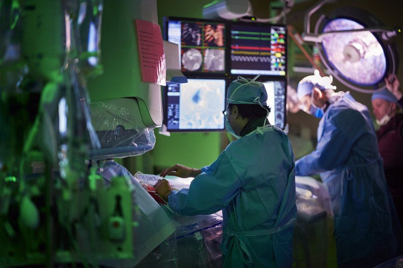
An interventional (IN-ter-VEN-shun-al) radiologist is a doctor with special training in performing image-guided procedures to diagnose and treat disease. These treatments are minimally invasive. That means they are done through just a small nick in the skin.
Methods that treat liver cancer include:
- Ablation
- Embolization
- Irreversible electroporation using a NanoKnife
- Laparoscopic and robotic surgeries
These methods target liver tumors without causing harm to other areas.
MSK also uses these methods to treat metastatic liver cancer.
Ablation for liver cancer
An interventional radiologist may do a percutaneous (per-kyoo-TAY-nee-us) ablation for liver cancer. Percutaneous (through the skin) ablation is a minimally invasive procedure that kills cancer cells without surgery.
Ablation for treating liver cancer targets only the tumor and a small margin of normal tissue right around the tumor. This method helps reduce side effects and the risk of the cancer coming back. You do not need to stay in the hospital to have this procedure.
We usually recommend ablation only if you have 3 or fewer liver tumors, each smaller than 3 cm in size.
There are a few types of ablation procedures for liver cancer. They kill cancer cells in different ways. Whether we choose heat, cold, or other methods depends on the size, location, and shape of the tumor.
What are the types of ablation for liver cancer?
Treating liver cancer with thermal (related to temperature) ablation
There are several kinds of thermal ablation.
- Radiofrequency ablation uses high-energy radio waves delivered by special needles placed right into the tumor. This kills the cancer cells with heat.
- Microwave ablation uses microwaves delivered by a special needle that creates high heat, killing the tumor.
- Cryoblation kills cancer cells by freezing them. This type of ablation is also called cryotherapy or cryosurgery.
- Other types of thermal ablation use lasers or focused ultrasound waves to kill tumor cells.
Treating liver cancer with chemical ablation
Chemical ablation kills liver tumor cells by injecting the tumor with a chemical, such as pure alcohol (ethanol).
Treating liver cancer with irreversible electroporation and a NanoKnife
It’s hard to treat some liver tumors with thermal (heating or cooling) ablation. That’s because they’re near bile ducts or blood vessels. Instead, your interventional radiologist may use a method called irreversible electroporation (ee-LEK-troh-por-AY-shun).
Irreversible electroporation (IRE) uses short electrical pulses to punch holes in cancer cell membranes. This damage to cancer cells kills the tumor.
The instrument used for this procedure is called a NanoKnife®. This method cannot treat as large an area as thermal ablation. But it can be used when a tumor is near a bile duct or blood vessel. It does not harm them.
What to expect during a liver ablation procedure
You will get anesthesia (medicine to make you sleep) before your ablation.
Your interventional radiologist will do this procedure through a small nick in your skin. They will put special needles through an incision (cut) in your skin. These needles deliver the treatment right to the tumor.
An ablation procedure often takes about 2 to 3 hours.
Your interventional radiologist will use imaging to guide the needle into the tumor. Imaging can include:
- Fluoroscopy (real-time X-rays)
- Computed tomography (CT) scan
- Magnetic resonance imaging (MRI) scan
- Ultrasound
Histotripsy
What is histotripsy treatment?
Histotripsy (HISS-toh-trip-see) treatment is a noninvasive way to break down cancer tumors using sound waves. Noninvasive (NON-in-VAY-siv) means it’s a procedure that does less harm to your body. This procedure is not surgery. It does not use knives or needles, heat or radiation.
How does histotripsy work to treat cancer tumors?
Histotripsy kills cancerous tumors by focusing ultrasound energy on the tumor cells. The energy creates tiny bubbles that grow fast and pop, breaking the tumor into tiny pieces. Your body then naturally clears away the dead cancer cells. Histotripsy only targets the tumor and a small area of tissue around the tumor. These areas are called margins. Histotripsy does not harm nearby healthy tissue, so it’s safer than some other treatment options.
Which cancers is histotripsy treatment used for?
Histotripsy is mainly used to treat liver cancers, such as hepatocellular carcinoma, cholangiocarcinoma, and metastasis. Metastasis happens when cancer cells travel to the liver from other parts of the body. Histotripsy is often used to treat cancer that spreads to the liver from the colon, breast, or pancreas.
Histotripsy may be a good option for people that cannot have surgery. It is used for tumors that are hard to remove with surgery or ablation. Ablation is a treatment that kills cancerous tumors using heat, cold, or other energy. These tumors are in areas of the liver where surgery or other treatment may be risky. Histotripsy can also be an option for people who want a treatment with fewer side effects.
Researchers are studying whether histotripsy is an option to treat other types of cancer, such as kidney and pancreatic cancers.
Histotripsy side effects and recovery
Histotripsy has very few side effects. It’s a cancer treatment that’s gentler on your body.
Since histotripsy does not use heat or radiation, it’s less likely to harm healthy tissue around the tumor. Most people have little or no pain after treatment. They can get back to their usual activities fast. Recovery time is much shorter than recovery from surgery or ablation.
Embolization for liver cancer
Arterial embolization (ar-TEER-ee-ul EM-boh-lih-ZAY-shun) is a procedure to block or limit arterial blood flow to a tumor in the liver. An artery is a blood vessel that carries blood from the heart to tissues and organs.
Like any tissue, tumors need oxygen and nutrients. Blocking the blood supply can kill tumor cells.
There are 2 blood vessels that send blood to the liver to feed it.
- The hepatic artery is the main source of blood for most primary liver cancers. This blood vessel supplies these tumors with blood that has the oxygen they need to survive.
- The portal vein carries blood to your liver from the intestines. It mostly feeds the part of the liver that does not have a tumor. It will keep feeding this part of the liver even after embolization blocks the hepatic artery.
Embolization may be an option for people who have liver tumors too large or in a hard spot for an ablation procedure.
What are the types of embolization for liver cancer?
There are a few ways to do a liver embolization procedure.
Bland transarterial embolization (tranz-ar-TEER-ee-ul EM-boh-lih-ZAY-shun), or TAE, cuts off or limits the blood supply to the liver tumor. Bland transarterial embolization does not use chemotherapy drugs to kill cancer cells.
Your interventional radiologist will thread a small catheter (thin flexible tube) into your hepatic artery. Then, they will inject (put) tiny beads in the catheter. These beads will kill tumor cells by blocking blood flow to the tumor.
There are 2 common tumor types that get most of their blood supply from the hepatic artery. They are primary liver tumors and neuroendocrine (nur-oh-EN-duh-krin) tumors. Bland embolization can work very well with these types of tumors.
Most people will have a fever or some pain. You may throw up or feel like you’re going to throw up for a few days after the procedure.
Embolization can be done again to treat cancer that recurs (comes back) after treatment. Embolization does not cure liver cancer, but it can kill most tumor cells in the area.
After embolization, your care team will monitor you very closely. You will have CT or MRI scans 1 to 2 months after the procedure. Then, you will have them every 3 to 6 months, for the rest of your life. We can treat the cancer again if it grows back.
Radioembolization (RAY-dee-oh-EM-boh-lih-ZAY-shun) is also knows as selective internal radiation therapy (SIRT). It’s a type of radiation therapy that treats liver cancer.
Your interventional radiologist will thread a small catheter (thin flexible tube) into your hepatic artery. Then, they will inject (put) tiny beads in the catheter that are loaded with radiation (yttirum-90, or Y90). They go into the artery that supplies blood to the tumors.
This method delivers internal radiation therapy right to the liver tumor over a few days. Because the beads are very close to the tumors, the radiation only affects the area near the tumor.
Radioembolization can be used to treat primary liver cancer.
Transarterial chemoembolization (tranz-ar-TEER-ee-ul KEE-moh-EM-boh-lih-ZAY-shun), or TACE, uses a mixture of chemotherapy drugs. They’re injected into the arteries that supply blood to the tumor. This is followed by small particles that block the blood supply to to keep the chemotherapy in the tumor.
MSK does not use TACE for liver cancer very often. Adding chemotherapy does not improve treatment results compared with bland embolization. The added drugs can also cause side effects and scar the arteries. That makes it harder to treat the tumors again, if needed.
Drug-eluting bead chemoembolization is another type of chemoembolization for liver cancer. In this procedure, the tiny beads used to block the blood supply are filled with chemotherapy drugs. The beads slowly release the drugs to kill the cancer cells.
We’re available 24 hours a day, 7 days a week