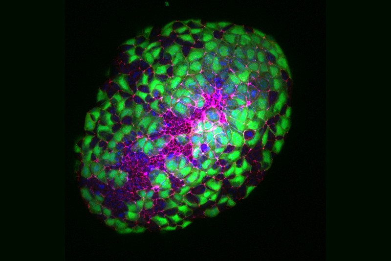
The Hadjantonakis Lab uses 3-D images like this one to learn how cells communicate with one another and how this communication contributes to the formation of various organs. By looking at embryos of different ages, they can learn how cells move around as the embryos develop.
The magenta shows a protein called ZO-1, which collects at the edge of cells to connect them with their direct neighbors and position them properly within the tissue. The green indicates a type of cell called the visceral endoderm, which helps embryos develop their structure. This includes formation of the notochord, which enables development of the central nervous system.
By studying the formation of mouse embryos, developmental biologists are able to learn more about how all mammals develop, including humans. Studying development contributes to our understanding of cancer because many of the processes that enable embryonic cells to specialize into more specific types are the same processes that go awry when normal cells turn into cancer.
Learn more about the areas of cancer research we’re most excited about.


