Skin cancer is the most commonly diagnosed cancer in the world, outnumbering all other types combined. This includes lower-risk skin cancer, such as basal cell carcinoma or squamous cell carcinoma, as well as the more serious melanoma. Most of these cancers are highly treatable, especially if discovered early.
Early detection can be challenging, however. It is often difficult for a person doing self-exams at home to tell if a suspicious spot is growing or changing. Traditionally, dermatologists take a biopsy (a sample of the tissue) for examination under a microscope by a pathologist.
Thanks to recent advances in imaging technology, Memorial Sloan Kettering’s Dermatology Service can now offer noninvasive approaches. These methods help in detecting, diagnosing, and even treating skin cancer.
“We’re one of the few centers in the world to have a comprehensive imaging program like this,” says dermatologist Anthony Rossi. “We are using the latest noninvasive imaging tools to detect these cancers and recurrences earlier, sometimes combining the technologies in novel ways.”
Scanning with Lasers to Diagnose Skin Cancer
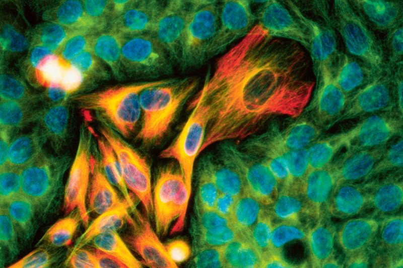
One new tool is reflectance confocal microscopy (RCM). This technology uses a low-power laser to scan skin lesions. The laser can penetrate slightly below the skin’s surface. The natural reflectivity of different cellular elements in a lesion provides important clues about whether it is cancerous or not. The reflected information can be collected in still and video images in microscopic detail.
“We can send these images electronically for examination and diagnosis by our doctors,” Dr. Rossi says. “In many cases, it eliminates the need for a conventional biopsy. Anything that does look suspicious can be followed up with a biopsy to confirm a cancer diagnosis.”
Since RCM is noninvasive, this method allows doctors to reexamine the same area repeatedly over time without hurting or changing the tissue, he explains. By contrast, taking several biopsies can damage tissue, and scarring can make it harder to study later.
Dr. Rossi says that MSK dermatologists are exploring the use of RCM in combination with another imaging technique called optical coherence tomography (OCT). This tool works at a greater depth than RCM — up to 1.5 millimeters — although the clarity is limited. By combining RCM with OCT, dermatologists obtain a fuller view of a lesion without cutting the skin.
RCM also provides guidance for surgery to remove skin cancers on the face and neck.
It is critical that surgeons can clearly delineate a tumor’s borders during a procedure. With RCM, MSK surgeons can image a tumor and its outer edges, what is called the margin, before an operation. Seeing the extent of a tumor more clearly enables surgeons to counsel patients and plan reconstruction. It also allows surgeons to be more precise when removing the cancer and spare as much normal tissue as possible.
The standard method of analyzing tissue requires a surgeon to remove a sample to be examined under a microscope. This can take several days. If cancer cells are found at the margin, a second surgery might be necessary. By using noninvasive imaging tools, MSK dermatologists are hoping to create a more streamlined approach, whether surgical or nonsurgical.
3-D Photography
MSK is one of a few centers in the world offering 3-D total-body photography. This technology can be used to track changes in the appearance of moles or lesions in people at a high risk of melanoma.
For this approach, a patient stands in the middle of a kiosk that houses 46 digital cameras. The cameras take photos simultaneously, and within a few minutes, a computer uses specialized software to process and assemble the images into a 3-D avatar. This digital model of the person’s body shows all the lesions, which doctors can zoom in on for closer inspection.
The approach creates a baseline record of a person’s entire skin surface that dermatologists can refer to during exams. The 3-D screening is available through MSK’s Melanoma Screening and Surveillance Program at the 60th Street Outpatient Center in Manhattan, as well at MSK’s New York regional sites in West Harrison, in Westchester County, and in Hauppauge, on Long Island.
Skin Cancer Imaging and Treatment Combined
For people with lower-risk skin cancers, such as superficial basal cell carcinoma or in situ squamous cell carcinoma, noninvasive imaging can guide nonsurgical removal of lesions. Dr. Rossi explains that he is treating these cancers with laser ablation or topical medications that can destroy the tumors with potentially less scarring and recovery time.
One limitation so far is that it is difficult to tell whether some of the cancer remains, so Dr. Rossi is investigating the use of laser ablation that is guided by RCM imaging to ensure that the skin cancer cells have been removed. If this approach proves effective, it would allow even more people to avoid surgery.


