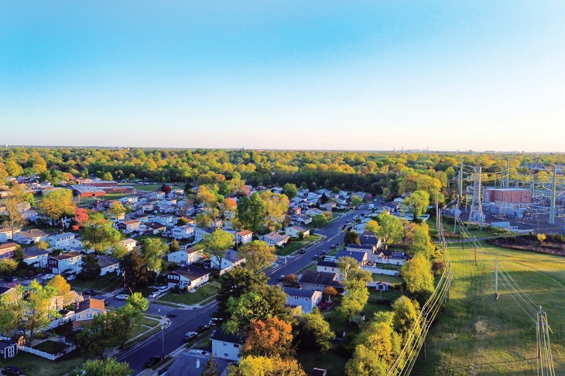
A neighborhood can change one loss at a time. A cornerstone business closes. A family moves away. Another falls on hard times. Soon the informal networks that hold a community together — the mutual support and watching out for one another — begin to fray.
But things don’t unravel all at once. They tip, gradually, as each small change reshapes conditions for everyone. And if those dynamics aren’t reversed, it can create a feedback loop of accelerating decline. Now scientists are coming to understand the ways in which something similar happens in the “neighborhood” surrounding cancer cells, which reshape their immediate environment to foster tumor development.
For decades, the field focused on the genetic changes inside individual cancer cells that cause them to go rogue. This enormously valuable work gave rise to tools like MSK-IMPACT®, which can identify the specific mutations driving a patient’s cancer and match them to drugs designed to target those mutations.
But a deeper puzzle remains: Genetic mutations alone are generally not enough to cause cancer. By the time we reach our senior years, we each carry an estimated 100 billion cells with cancer-causing mutations — yet many people never develop cancer. That’s because the environment around a mutation-carrying cell also plays a critical role in what happens next.
Cancer cells, researchers are finding, are active architects of their environment — sending out signals, corrupting neighboring cells, weakening the immune response, feeding off and causing inflammation, and remaking the neighborhood in their own image.
Understanding this process — and learning to interrupt it — is the goal of an emerging field of research that sees cancer not just as a disease of individual cells gone bad, but also of the compromised neighborhoods around them. MSK is helping to lead this evolution in thinking, fueled by a transformative philanthropic gift that created the Marie-Josée and Henry R. Kravis Cancer Ecosystems Project.
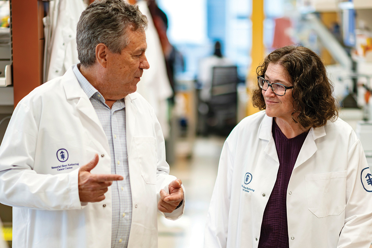
“Cancer cells don’t exist in isolation,” says cancer biologist Scott Lowe, PhD, the project’s scientific director and Chair of MSK’s Cancer Biology and Genetics Program. “From the very beginning, they’re engaging in complex interactions with their neighbors and with the immune system.
“What we’re learning,” he adds, “points to widespread features of cancer neighborhoods that transcend individual cancer types — features that are already paving the way for new prevention and treatment strategies.”
Cancer: A wound that doesn’t heal
The understanding of tumors as “wounds that do not heal” has guided medicine for decades. Now MSK researchers are showing in remarkable detail just how cancer hijacks the programs that repair injuries — and how it spreads its influence to its neighbors.
Consider what happens when you slice your finger. Within moments, the surrounding tissue springs into action: It sends signals that there’s an injury, immune cells rush in to ward off infection, new skin cells are generated to close the cut. And then, critically, when the job is done, the alarm is switched off.
Cancer borrows this same emergency response, but the repair never completes. The alarm never stops, and the tissue remains in a state of crisis.
When flexible cells have faulty brakes
A recent collaboration between Dr. Lowe and computational biologist Dana Pe’er, PhD, revealed how this hijacking works and how it’s driven by a surprisingly small number of “highly plastic” cells.
These are cells that respond to injury by temporarily becoming unusually flexible, so they can produce whatever new cells are required to make the repair. But when cancer-causing mutations are present and when the genes that normally prevent tumors from growing are also compromised, that temporary emergency can extend indefinitely.
Among the genes that suppress tumors, p53 is the most famous and well-studied, earning it the nickname “guardian of the genome.” The genomes of cells that lose p53 become unstable and develop further changes that can fuel cancer progression.
The MSK study revealed that the tug-of-war between p53 and cancer-driving programs takes place in these highly flexible, plastic cells: When p53 is turned on, it acts as an emergency brake on cancer progression — and when it’s lost, the cancer can prevail.
“What we’re finding is that p53 is even more than the guardian of the genome — it’s the guardian of plasticity,” says Dr. Pe’er, Chair of MSK’s Computational and Systems Biology Program and a Howard Hughes Medical Institute (HHMI) Investigator. “Cells need to become flexible for healing and repair to happen, but p53 is there to pump the brakes if they try to go rogue. Without p53, this flexibility can be hijacked by cancer.”
The advanced computational and artificial intelligence–driven approaches developed in recent years by Dr. Pe’er’s lab, in conjunction with MSK’s Single-Cell Analytics Innovation Lab (SAIL), have allowed MSK researchers to push the boundaries of what spatial imaging technologies and cell-by-cell analyses can teach us — enabling the study of cancer neighborhoods in unprecedented granularity.
“Most artificial intelligence is task-oriented, optimized to solve one particular type of problem,” Dr. Pe’er says. “We treat it more like a microscope, a true discovery tool.”
A few bad apples
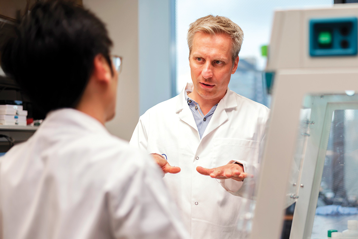
It doesn’t take many of these abnormal, highly plastic cells without p53 to cause havoc, according to a related study from the lab of Tuomas Tammela, MD, PhD. Working in mouse models of lung cancer, his team found that this population makes up only about 3% of cells in early lesions — yet drives cancer’s ability to grow, resist treatment, and advance. As disease progresses, their numbers rise to about 15% in established tumors and up to 30% in cancers that have spread.
“But if we kill off these plastic cells very early, we can prevent mutated cells from ever becoming cancers, our research shows,” Dr. Tammela says, noting the work could inform prevention strategies in high-risk populations, such as smokers.
Dr. Lowe’s lab made a similar finding in a mouse model of pancreatic cancer. Almost all pancreatic cancers are driven by KRAS mutations. A short course of treatment with a KRAS-blocking drug was enough to disrupt the early-stage cancer neighborhood.
“A three-day treatment in mice paused cancer development for months, which would be the equivalent of years on a human timescale,” says Dr. Lowe, who is also an HHMI Investigator.
How cancer corrupts its neighborhood
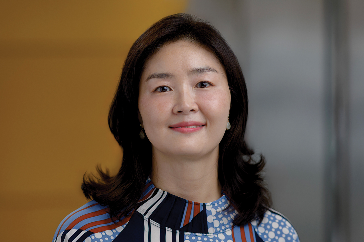
When cells get stuck in emergency repair mode, the damage doesn’t stay contained — it spreads to nearby tissue and immune cells. A study from the lab of developmental biologist Joo-Hyeon Lee, PhD, tracked individual lung cells from the moment they acquired cancer-causing mutations, mapping in mouse models and lab-grown miniature organs how neighborhood corruption begins even before a tumor forms.
The research showed the mutated cells broadcast distress signals to surrounding tissue, triggering the formation of scar tissue and recruiting immune cells that suppress the body’s defenses rather than attacking the cancer — essentially adapting the surrounding area to support a tumor. When the team blocked those initial distress signals using a lung cancer drug, these changes were prevented. And where tumors had already started to form, many reversed course.
“Communication with nearby healthy cells to build a ‘pre-cancer niche’ is essential for tumors to grow,” Dr. Lee says. “Encouragingly, at these early stages, it’s possible to switch those altered surrounding cells back to normal.”
Cleaning up the neighborhood
Together, these studies and others from across MSK are not only shedding new light on how cancer ecosystems develop, function, and reinforce themselves — they also point directly toward new treatment strategies.
For example, another recent study from the Lowe Lab showed that a type of personalized immunotherapy called CAR T cell therapy could be used to target cancer neighborhoods.
The team, in collaboration with researchers at Columbia University, identified a protein called uPAR that is found on the surface of tumor cells, and on the surrounding tissue and immune-suppressing cells that help cancer thrive.
Critically, however, uPAR is not found on most healthy cells. And when researchers targeted uPAR in mouse models in the lab, it dramatically shrank lung, pancreatic, and ovarian cancers — and even cleared metastases in some experiments.
Based on these encouraging results, MSK researchers are now working to advance uPAR-targeting therapeutics into clinical trials.
“Our findings point toward a new era where we don’t just go after tumors, but also the broken neighborhood that supports cancer,” Dr. Lowe says. “The MSK Ecosystems Project is bringing together researchers across labs and disciplines to investigate these key ideas from a variety of angles.
“And the patterns we’re uncovering appear true across different types of cancer,” he adds. “That gives us real hope that therapies targeting these neighborhoods could work for many patients — whether the goal is cancer prevention, early interception, or treating advanced disease.”
Video: Bold science to target cancer ecosystems
Bad News for Cancer: New Technique Shrinks Solid Tumors in the Lab
Dr. Lowe’s research is supported by the MSK donor community, including Break Through Cancer, The Mark Foundation for Cancer Research, and The Lustgarten Foundation for Pancreatic Cancer Research.
Dr. Pe’er’s research is supported by the MSK donor community, including Alan, Sandra, and Adam Gerry through their Gerry Foundation.
Dr. Tammela’s research is supported by the MSK donor community, including The Mark Foundation for Cancer Research.
The Marie-Josée and Henry R. Kravis Cancer Ecosystems Project is supported by the MSK donor community, including The Marie-Josée and Henry R. Kravis Foundation.
Dr. Lowe holds a Geoffrey Beene Cancer Research Center Chair and is a Howard Hughes Medical Institute Investigator.
Dr. Pe’er holds the Alan and Sandra Gerry Endowed Chair and is a Howard Hughes Medical Institute Investigator.