Summary of Invention
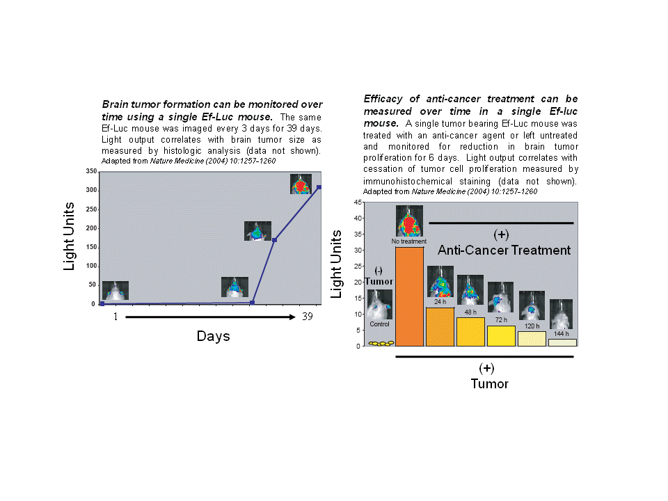
The Ef-Luc mouse is a transgenic mouse model expressing luciferase driven by an E2F1 responsive promoter used for the sensitive, non-invasive, in vivo detection of tumor growth. Tumor formation as well as efficacy of anticancer treatment can be monitored over time using a single Ef-Luc Mouse.
The target market for the Ef-Luc mouse is preclinical CROs, biotech and pharma research and development operations, and academic researchers.
Background
E2F1 is a transcription factor whose activity is repressed by the retinoblastoma protein (Rb), a master regulator of cell-cycle progression through the G1 to S transition. A common feature in many distinct types of human malignancies is the loss of Rb function, resulting in upregulation of E2F1 transcriptional activity and dysregulation of cell-cycle control. Therefore, the Ef-Luc mouse can be considered a general reporter animal useful for the detection and imaging of multiple different tumor types.
The Ef-Luc mouse is an ideal tool for monitoring cell-cycle activity during tumor development in a living animal using bioluminescence imaging. Areas of abnormally high cell proliferation in the Ef-Luc mouse, namely cancerous cells, drive expression of luciferase. The resulting luciferase can be detected by injection of the Ef-Luc mouse with the luciferase substrate luciferin; luciferase oxidization of luciferin produces light that is then detected through the body of the mouse and is proportional to tumor cell burden.
Advantages
- High sensitivity allows detection of small subcutaneous tumors (<1,000 cancer cells) and deeper lesions (1-3 cm deep), which can be undetectable by standard measurement methods.
- Universal tumor detection increases the applicability of the Ef-Luc mouse model to multiple tumor types.
- Quantitative measurement of tumor burden reveals subtle changes in tumor growth.
- Rapid real-time imaging allows spatial and temporal resolution of tumor growth.
- This noninvasive method with minimal toxicity allows repeated imaging of a single animal.
- Fewer mice are needed per study, which reduces the cost of animal studies.
Areas of Application
- Efficacy evaluation of anticancer treatments and therapies
- Assessment of carcinogenic potential of compounds and environmental insults
- Development of novel bioluminescent models of known cancer mouse models by cross-breeding
- Evaluation of metastatic potential of primary tumors
- Investigation of molecular mechanisms critical for tumor maintenance
References
Uhrbom L, et al. (2004) Nature Medicine. Nov; 10(11):1257-1260.
Lead Inventor
Eric C. Holland, MD, PhD
Patent Information
U.S. patent issued: 7, 041, 869
