Hedgehog proteins are some of the most important molecules in biology. These proteins help regulate cell growth, specialization, and patterning in the development of embryos by acting as messengers between cells. In addition to playing a key role in how proper tissues are formed, abnormal Hedgehog messaging has been linked to several types of cancer, including certain pancreatic cancers.
Before Hedgehog proteins can function as messengers, however, they need to be assembled. Scientists in the Sloan Kettering Institute, led by structural biologist Stephen Long, published a paper in Science on June 10, 2021, that reveals how one of the “assembly machines” functions.
“We’re interested in understanding how this assembly works on a detailed atomic level,” says Dr. Long, a member of SKI’s Structural Biology Program. “We know that some cancers use Hedgehog to drive uncontrolled cell growth, and a better understanding of its assembly process may lead to new treatment options.”
Watching an Enzyme Do Its Work
Technology Aids in Important Discovery
One enzyme that’s crucial to the assembly of Hedgehog proteins is known as Hedgehog acetyltransferase, or HHAT. It acts like a machine on an assembly line to link two components together and form the final Hedgehog product. Once HHAT completes this assembly step, the finished Hedgehog protein acts as a messenger. For this reason, inhibitors of HHAT, which prevent it from completing its assembly, could potentially be useful for the treatment of certain cancers that depend on Hedgehog messages.
MSK scientists are able to probe the shapes of proteins more completely than ever before thanks to an advanced imaging technology called cryogenic electron microscopy (cryo-EM), which MSK acquired in 2016.
Older methods of looking at proteins’ structures, such as X-ray crystallography, require molecules to be crystallized in a repeating array, similar to a salt crystal. This can limit the ability to study proteins that are flexible. For the HHAT enzyme, it’s particularly challenging because the protein is normally embedded within a membrane inside a cell.
“Membrane proteins are generally more difficult to study in the laboratory than other types of proteins, and, consequently, we know less about how they work,” Dr. Long says. Because cryo-EM doesn’t require proteins to be crystallized, researchers can now decipher what the proteins look like more easily.
Watching an Enzyme Do Its Work
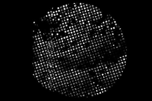
Thanks to the cryo-EM, Dr. Long and his colleagues were able to visualize how HHAT creates that final linkage between the Hedgehog precursor protein and a lipid (fat) called palmitate. Using the high-powered microscope, the researchers were able to take millions of pictures of individual HHAT molecules. These pictures revealed information similar to that obtained from X-rays of an arm.
After careful computational analysis of the pictures, the researchers were able to deduce the detailed atomic structure of HHAT — essentially what it would look like if you could see it up close in three dimensions. They were also able to visualize the enzyme in the process of completing the linkage step. By inspecting this process, they were then able to hypothesize how HHAT forms the linkage and test those hypotheses using other experimental approaches.
What they discovered was particularly intriguing. HHAT is relatively unusual for an enzyme in that it resides within a cellular membrane (the membrane of an organelle called the endoplasmic reticulum). The researchers discovered that HHAT obtains the palmitate component from one side of the membrane and the Hedgehog component from the other side of the membrane. The enzyme links these two parts together within the membrane and then releases the product toward one side. In this way, the completed Hedgehog protein emerges from the enzyme like an automobile rolling off the assembly line.
Dr. Long credits two of MSK’s core facilities — the Structural Biology Core and the Antibody and Bioresource Core — that were vital to this research. Antibodies to HHAT were important because it made HHAT easier to recognize in the cryo-EM pictures. These pictures were collected using the cutting-edge microscope in the Structural Biology Core. Yiyang Jiang, a postdoctoral fellow in the Long lab, was the study’s first author. “None of this would have been possible without Yiyang,” Dr. Long says. “He carried out nearly every aspect of this project.”
Basic Findings with Broad Implications
Agents that target the activity of HHAT that could be developed as potential cancer therapies are already being studied by SKI cell biologist Marilyn Resh. Dr. Long and Dr. Resh are now collaborating to learn more about how these agents work and ways they could be optimized for the development of cancer drugs.
One goal is to use the cryo-EM to visualize how these potential drugs interact with HHAT and prevent it from linking palmitate to Hedgehog proteins.
“With this research we have discovered some of the fundamental biophysics and biochemistry about how the HHAT enzyme works,” Dr. Long says. “We hope that it will lead to new discoveries for cancer treatment. It also helps us learn about the activity of related enzymes that control other important cellular processes.”


