Seven students will be awarded PhD degrees on May 19, 2021, from the Gerstner Sloan Kettering Graduate School of Biomedical Sciences (GSK), an innovative doctoral program that prepares the next generation of basic laboratory scientists to work in research areas related to human disease with a focus on cancer. The virtual commencement ceremony, which will take place as part of Memorial Sloan Kettering’s 42nd annual academic convocation, brings the total number of GSK’s alumni to 82.
“I’m very proud of this gifted, hard-working group of students and look forward to celebrating their impressive academic achievements,” says GSK Dean Michael Overholtzer.
This year’s graduation will feature a special commencement address from Howard Hughes Medical Institute Investigator Joan A. Steitz, Sterling Professor of Molecular Biophysics and Biochemistry at Yale School of Medicine. A leader in the field of microbiology, Dr. Steitz has been recognized for her pioneering role in expanding our understanding of RNA biology and for her lifelong advocacy for inclusion of women in the sciences. Among her numerous accolades, she was awarded the prestigious Wolf Prize in Medicine in 2021 and the 2018 Lasker~Koshland Special Achievement Award in Medical Science, one of the highest forms of recognition that a scientist can receive.
“We are thrilled that Dr. Steitz will address our students during GSK’s 10th commencement,” says Dr. Overholtzer. “Her remarkable body of work and professional insights are sure to inspire our graduating scientists as they ambitiously embark on their promising careers.”
GSK will award doctorates to Tyler Hitchman, Brian Joseph, Xiaoyi Li, Michelle M. Riegman, Ryan Smith, Benjamin Tischler, and Zeda Zhang. Throughout the past year, the students successfully defended their dissertations, the final requirement in earning their doctorates. Their thesis research reflects GSK’s mission to train its students to tackle real-life clinical challenges through biomedical investigation. Learn more about their investigations below.
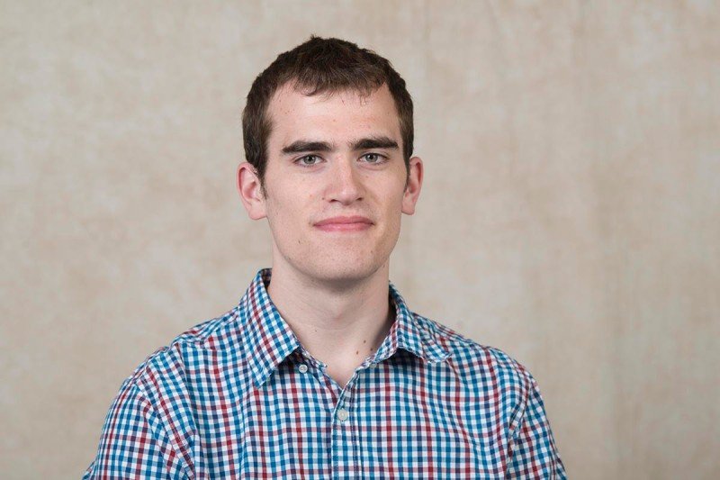
I completed my thesis in the laboratory of physician-scientist Yu Chen, who focuses on understanding the role of transcription factors and other proteins that drive oncogenesis in a variety of cancer types. My research centered on uveal melanoma (UVM), a rare eye cancer that lacks approved therapies for metastatic disease. My work investigated pathways essential for the development of the disease and shed light on how to effectively target it.
UVM, and a fraction of other melanoma subtypes, are driven by genetic mutations in the Gαq pathway. We developed models for these driver mutations, including rarer alterations that are not readily available to study. We found that all mutants were highly and selectively sensitive to treatment with a natural compound that directly targets Gαq. Furthermore, this compound inhibited cell growth and signaling in human UVM cells as well as tumor growth in xenograft experiments.
Using transcriptomic data, we also identified that the MAPK pathway quickly rebounded after treatment, indicating potential resistance mechanisms to the compound. We developed a combination therapy using a MEK inhibitor that resulted in sustained long-term inhibition of the MAPK pathway and led to significant reductions in tumor volume and even tumor shrinkage in both cellular and mouse models of UVM.
Our findings, which are published in Clinical Cancer Research, indicate that the targeted inhibition of the Gαq pathway alone and in combination with MEK inhibition provides a promising therapeutic strategy for UVM.
I chose to pursue my PhD at GSK because it offers small class sizes and the opportunity to learn and conduct translational cancer research within an intimate community of peers and faculty. I am a previous recipient of the Ruth L. Kirschstein National Research Service Award (NRSA) Individual Predoctoral Fellowship and am now a postdoctoral research fellow at MSK.
In 2012, I participated in the Summer Undergraduate Research Program (SURP) at GSK in the laboratory of Daniel Heller. This was the backdrop to a truly remarkable summer, during which I witnessed a vibrant and collaborative research environment and got the chance to appreciate both MSK’s mission and the pace of life in New York City. The experience cemented my desire to pursue a PhD in the biological sciences and also paved the way to do so at GSK.
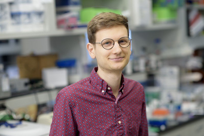
I completed my dissertation under the mentorship of developmental biologist Eric Lai. His laboratory focuses on comprehending how complex biological patterns can be precisely assembled to execute appropriate cellular behaviors at the right times, in the right places, and in the right numbers.
RNA is involved — directly and indirectly — in all biological processes. I was drawn to Dr. Lai’s work studying transcriptional regulation through the lens of RNA metabolism pathways to understand how unusual RNA processing events contribute to animal development. My thesis project used a combination of genetics, molecular biology, and bioinformatics to understand the complex properties of cryptic splice sites (CSS) during RNA splicing in the fruit fly.
Precursor-mRNAs (pre-mRNA) undergo several processing events prior to becoming messenger RNAs (mRNA) that make proteins. One essential step is RNA splicing, in which protein coding sequences (exons) are stitched together and intervening sequences (introns) are removed. This highly precise process is guided by the activity of stereotypic splice sites that mark the boundaries of exons and introns. Interestingly, the pre-mRNA landscape also contains thousands of CSS that match splice motifs but are not directly implicated in the splicing.
I explored the functions of these elements during RNA processing by studying recursive splicing (RS). RS is a mechanism in which a long intron is removed as two or more smaller, manageable fragments. When I began my thesis research, our only insights into this process were atypical splice substrates called recursive splice sites. Data indicated that these sites could only be found on incredibly long pre-mRNAs, but we knew virtually nothing about their activity.
I manipulated these elements in the fruit fly and in cell cultures and studied their sequence patterns to derive several interesting ideas about CSS and splicing. First, I found that recursive splice sites are not solo actors; instead, they function as short exons by utilizing cryptic splice sites that are downstream. I also discovered that these short cryptic exons are part of a continuum that includes annotated expressed exons. These findings revised the phenomenon of recursive splicing as a mechanism of alternative splicing.
In addition, I uncovered that hundreds of CSS — including recursive splice sites — on canonical exons are actively suppressed by the exon junction complex (EJC). The EJC is deposited onto mRNAs during intron removal. My work indicates that this event increases the fidelity of splicing in part by blocking the detection of CSS.
These discoveries, which are published in Nature Structural & Molecular Biology and have been accepted for publication in PLOS Genetics, underscore the ever-expanding list of functions of CSS during splicing and the opportunity for further investigation.
I am a previous recipient of the Chairman’s Prize at MSK and am now a postdoctoral fellow in Kevin Eggan’s lab at Harvard University in Cambridge, Massachusetts, where I am studying altered RNA metabolism in motor neuron disorders.

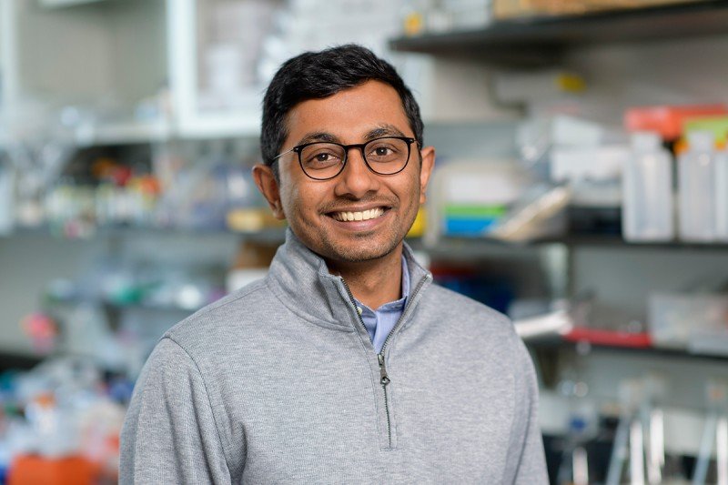
I completed my dissertation research in the laboratory of cancer biologist Andrea Ventura. His lab focuses on understanding microRNAs, which play important roles in mammalian development and, when disrupted, in disease. At the molecular level, microRNAs exert their functions by recognizing and binding to messenger RNAs and tuning their levels of expression in cells.
MicroRNA research has been stymied by difficulties in determining the direct biological targets of microRNAs. To overcome this obstacle, we developed a new tool that simplifies microRNA target mapping in cells and live tissues in mice. We applied the method, called Halo-enhanced Ago2 pulldown (HEAP), in a variety of biological contexts, including mouse embryonic development and tumor formation.
As a result, we were able to generate high-resolution maps that delineated microRNA-target interactions in cells. Our analysis of these maps revealed unique microRNA regulation signatures in each context. By comparing the maps obtained from tumors and normal tissues, we identified microRNA regulation events that may be crucial for cancer development.
We hope that the method we developed will serve as a resource to assist the scientific community in conducting functional studies of this important class of non-coding RNAs in the in vivo setting. The results of our work are published in Molecular Cell.
I chose to pursue my doctorate at GSK because of the high-quality research conducted at MSK. I am a previous recipient of the Geoffrey Beene Graduate Student Fellowship at MSK and am currently a postdoctoral fellow at the University of Washington.
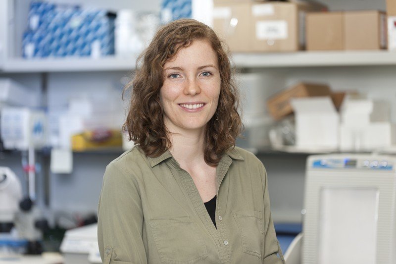
I completed my thesis work in the laboratory of GSK Dean Michael Overholtzer. His research centers on learning about the mechanisms of nutrient sensing, cellular responses to nutrient starvation, and processes of apoptosis, a type of cell death that plays an important role in many human diseases, including cancer.
When cells die, they can do so through many different mechanisms, each with their own triggers, molecular pathways, and cellular outcomes. Each route to a cell’s death can have a different effect on its surrounding cells. My thesis focused on ferroptosis — cell death by iron that is linked to a cell’s use of oxygen for metabolism.
When we induced ferroptosis in clusters of cells, we noticed that the death seemed to spread outward from the first dying cell, like falling dominoes, resulting in a wave of death traveling across the cell population. We had never seen anything like this occur with other forms of cell death, so we wondered whether there was something special about ferroptosis that caused dying cells to push their neighbors closer to death.
To test this, we teamed up with computational biologists at the Ben-Gurion University of the Negev in Beer-Sheva, Israel, to develop a method that would allow us to quantify the spatiotemporal dynamics of different forms of cell death. We showed that there is indeed a clear pattern to the cell death observed during ferroptosis and a strong correlation with neighboring cells’ deaths that was absent in other types of cell death. This suggests that ferroptosis is able to spread from cell to cell.
Other researchers have discovered that ferroptosis is partially responsible for the tissue damage observed after an ischemic heart attack or stroke, which often presents as large continuous zones of necrotic cells. It may therefore be possible that the wave of death we observed in the petri dish also occurs when ferroptosis is triggered in the human body. Understanding how this death spreads may help us limit the amount of tissue damage that occurs.
Because many types of cancer are thought to be particularly sensitive to ferroptosis, several ferroptosis-inducing therapeutics are currently being developed. Interestingly, we also found that not all of these drugs trigger the wave of death, so it may be important to focus development efforts on the agents that do because these could potentially eliminate larger numbers of cancer cells in one pass compared to therapies that induce other, non-spreading forms of cell death.
The results of our work are published in Nature Cell Biology.
I am a previous recipient of the Grayer Fellowship and the Robert B. Catell Fellowship at MSK. I am now a manager in Technical Regulatory Affairs for the Marketed Biologics Group at Roche.
I completed my dissertation under the mentorship of neurosurgeon-scientist Viviane Tabar, whose research focuses on utilizing human stem cells as regenerative tools for brain repair and for studying brain cancer. My thesis project investigated how different subtypes of stem cells in the brain respond differently to mutations that are thought to cause brain tumors in children.
Pediatric brain tumors occur in very specific patterns of where they emerge in the brain and when they are diagnosed during the course of childhood. These patterns suggest that there are specific areas of the brain during development that might be more susceptible than others to having a tumor form as an adverse effect of abnormal tissue growth.
Neural stem cells are the cells that build the brain by giving rise to mature brain-cell types like neurons and glial cells. They are present across the developing brain but might have inherent differences over space and time that would explain why tumors grow in some locations and time frames more than in others.
My project centered around the question of whether different neural stem cell subtypes respond differently to one particular genetic alteration called histone H3.3 K27M, which is found in pediatric brainstem tumors of early to middle childhood.
To test this, I generated new techniques to derive neural stem cell subtypes, representing different brain regions and cellular stages of maturity, from human pluripotent stem cells. I then asked how each of these subtypes responded when I introduced H3.3 K27M into their DNA, with the hypothesis that cells susceptible to forming a tumor would grow more with the mutation than without it.
I found that, while neural stem cell subtypes from different areas of the brain all responded similarly to H3.3 K27M, there was evidence of a time-dependent effect on the cells’ susceptibility to increased growth. More-mature cells that arose at a later stage of development did not grow more in response to the mutation, unlike their less-mature counterparts that emerged at an earlier stage.
These findings indicate that pediatric brain tumors with H3.3 K27M may require features of an early stage of neural stem cell development for tumor formation. These insights may provide clues into how researchers can gain a better understanding of this lethal type of childhood cancer and investigate new treatments for it.
The results of our work will be published soon.
Previously, I received the Ruth L. Kirschstein National Research Service Award from the National Cancer Institute as well as the Grayer Fellowship and the Geoffrey Beene Graduate Student Fellowship from MSK.
I was drawn to the GSK program because of its supportive community of students as well as the non-traditional academic structure that prepares students for a variety of career paths. I learned a great deal about stem cell biology and regenerative medicine and gained professional skills that have benefited me in my current role as a translational neuroscientist at BlueRock Therapeutics in New York.
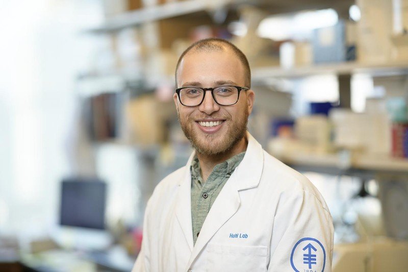
My interest in science is based on leveraging knowledge to improve patient outcomes. GSK allowed me to keep sight of this goal in my doctoral research while introducing me to areas of science I had not even considered when choosing a graduate school. While I assumed that I would pursue cancer biology, I ultimately chose to study antifungal immunology.
I completed my thesis research in the laboratory of physician-scientist Tobias Hohl, Chief of the Infectious Diseases Service at MSK. His lab studies human fungal pathogens and how the immune system responds to them.
My research focused on the role of platelets in host defense against a common fungus called Aspergillus fumigatus (A. fumigatus) and a severe fungal infection — invasive aspergillosis (IA) — that is caused by this pathogenic mold.
As part of its life cycle, A. fumigatus releases spores, called conidia, into the air. Normally, humans inhale hundreds to thousands of conidia daily. In healthy people, the cells of the innate immune response rapidly inactivate and clear the conidia. However, when the immune system is injured — such as in people undergoing cancer treatments like corticosteroids or chemotherapy — the conidia can escape the immune system, germinate into long strands called hyphae, cause tissue damage, and disseminate throughout the body.
Even with modern-day therapeutics, fungus-caused mortality can reach 25 percent in high-risk groups. With more than 200,000 global IA cases annually and a high financial burden on the medical system, a better understanding of this deadly disease and how the body responds to it is an urgent public health concern.
Thrombocytopenia (a low count of platelet cells that help blood clot) has been identified as a risk factor for IA. However, it often occurs at the same time as neutropenia, or a low level of neutrophils (a type of white blood cell that helps fight infection). Neutropenia is itself well appreciated as the primary risk factor for IA, making it challenging to study the clinical role of platelets in the immune response to Aspergillus.
My dissertation project utilized two mouse models of thrombocytopenia to test the role of platelets in preventing poor IA outcomes. I discovered that in the absence of platelets, mice were highly susceptible to Aspergillus infections. While platelets were involved in ensuring optimal early neutrophil activity, their primary role was to prevent severe bleeding into the lungs during the inflammatory response.
Further investigation is needed to understand whether platelets can form the basis of therapeutic strategies in people at risk for IA. The results of our work are published in PLoS Pathogens.
The opportunity to learn from my talented peers and a world-renowned faculty at GSK was an incredible experience. I previously received the Ruth L. Kirschstein National Research Service Award (NRSA) Individual Predoctoral Fellowship from the National Heart, Lung, and Blood Institute. I am currently a Life Sciences Specialist at L.E.K. Consulting in New York.
I conducted my thesis work under the guidance of Howard Hughes Medical Institute Investigator Charles Sawyers, Chair of MSK’s Human Oncology and Pathogenesis Program. His laboratory develops molecularly targeted approaches to treat cancer. I was particularly interested in his study of resistance to hormone therapy in prostate cancer.
My dissertation research shed light on the mechanisms of two distinct but related clinical characteristics that are commonly seen in men with prostate cancer. Understanding the molecular survival mechanisms behind both scenarios may facilitate the development of new therapies.
Patients with high-grade primary tumors rarely have a complete pathological response to even the most potent hormone therapy regimens prior to surgery. This incomplete response, in which evidence of disease remains, often drives disease relapse and drug resistance.
In the first project, I used column fractionation and purified a growth factor from the tumor microenvironment called neuregulin 1 (NRG1) that is responsible for resistance to hormone therapy in prostate cancer. NRG1 is secreted by cancer-associated fibroblasts and serves as a mechanism of drug resistance by activating HER3 in the tumor.
Interestingly, NRG1 expression is induced in some patients receiving hormone therapy, indicating an adaptive response to treatment. We found that blocking NRG1 resensitizes tumors to hormone deprivation in vivo and may explain why patients with high-grade prostate cancer rarely achieve complete pathological responses to hormone therapy. We also developed a clinical grade NRG1 immunohistochemistry assay as a biomarker with the aim of identifying patients with elevated NRG1-HER3 activity who might benefit from anti-NRG1/HER3 antibody therapy. These findings are published in Cancer Cell.
Another common characteristic among men with prostate cancer is that many metastatic tumors escape their dependencies on hormone signaling and acquire resistance to treatment. Metastatic prostate cancer is also characterized by recurrent genomic-copy number alterations that are presumed to contribute to resistance to hormone therapy.
In the second project, I performed an in vivo screen for genetic drivers of antiandrogen resistance and identified a gene called CHD1 as a factor known to play an important role in the stem cells’ ability to develop into different cell types. I found that loss of CHD1 causes the dysregulation of chromatin (material that makes up chromosomes) and facilitates the evolution of drug-resistant tumors by promoting a cell lineage identity to switch away from a luminal epithelial identity. This lineage plasticity is driven by four transcriptional factors, further emphasizing the need for combination therapy to target heterogeneous mechanisms of antiandrogen resistance in advanced disease. The results of this project are published in Cancer Cell.
Taken together, my dissertation research has elucidated two distinct mechanisms of resistance to hormone therapy in both primary and metastatic prostate cancer. These findings have not only expanded our conceptual knowledge of how drug resistance develops, but they have also provided actionable therapeutic strategies that may increase the efficacy of current therapies.
I was drawn to the GSK program because of the quality of MSK’s basic and translational research. I received cutting-edge training in a multidisciplinary, collaborative setting that has prepared me with the skills and knowledge necessary to conduct biomedical research with a direct impact on the clinical management of people with cancer. I am a previous recipient of the National Cancer Institute Predoctoral to Postdoctoral Fellow Transition Award and have chosen to stay at MSK as a postdoctoral research fellow in the laboratory of cancer biologist Scott Lowe, where I am studying human aging and cancer.