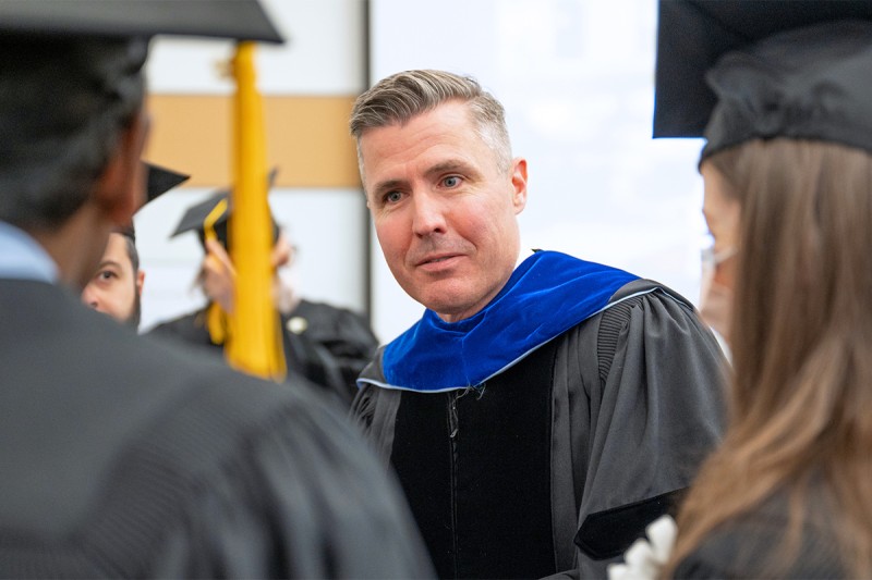
This year 17 scientists who did their training at the Gerstner Sloan Kettering Graduate School of Biomedical Sciences (GSK) are poised to receive their doctorates, the highest number ever to graduate from GSK in a single year. Their years of dedication and training will be recognized on May 17, 2023, when they will be awarded their PhD degrees from GSK. This year’s commencement marks the school’s 12th graduating class since admitting its first class of students in 2006.
GSK offers the next generation of biomedical investigators an intensive doctoral program in cancer biology. The program trains them to study and attack cancer through the twin lenses of basic research and real-life clinical challenges. The graduates will receive their diplomas as part of the 44th annual academic convocation ceremony at Memorial Sloan Kettering Cancer Center (MSK), bringing the total number of GSK alumni to 107.
“We are very pleased to recognize the contributions of this outstanding group of students,” says GSK Dean Michael Overholtzer, PhD. “Each of them has already made important discoveries that have contributed to our understanding of cancer and related fields. We are so excited to see what they all accomplish in the future.”
This year’s graduation ceremony will feature a special commencement address from Craig B. Thompson, MD, former President and CEO of MSK. Dr. Thompson led MSK for 12 years before stepping down to focus on his lab research. Dr. Thompson is widely recognized for his discoveries related to the genes that control programmed cell death and how the malfunction of such genes can contribute to cancer. More recently, his research has focused on the role that metabolic changes play in the origin and progression of cancer — work that has sparked renewed interest in the broader topic of cancer metabolism.
“This year we are so pleased to be able to honor our friend and colleague Craig Thompson,” Dr. Overholtzer says. “We are looking forward to recognizing all of his contributions to GSK and to Memorial Sloan Kettering more broadly.”
This year GSK will award doctorates to Paige Krystin Arnold, Bradley Benjamin, Tingxu Chen, Mollie Elizabeth Chipman, Anton Dobrin, Caroline Gleason, Florisela Herrejon Chavez, Nayan Jain, Yasemin Kaygusuz, Buren Li, Miguel Angel Miranda-Román, Stella V. Paffenholz, Gemma L. Regan-Mochrie, Adina Rachel Schonbrun, Maria Sirenko, Zhongmin Wang, and Adele Whaley. Throughout the past year, these students have successfully defended their dissertations, the final requirement in earning their doctorates. Learn more about their research projects below.

My thesis work centered on the observation that mammalian cells display significant heterogeneity in tricarboxylic acid (TCA) cycle behavior, both in normal cells and under pathological conditions like cancer. The TCA cycle — otherwise known as the Krebs cycle — is a core metabolic pathway that is responsible for several critical functions in growing cells, including energy production and the synthesis of cellular building blocks.
Despite the importance of TCA cycle-derived products for cell survival and growth, TCA cycle activity varies significantly across different cell types. The major goals of my dissertation research were to identify drivers of TCA cycle heterogeneity and determine whether TCA cycle behavior is crucial for the establishment of cell identity.
By studying TCA cycle activity using carbon tracing techniques, we uncovered an alternative, “noncanonical” TCA cycle active in both normal cells and cancer cells. Using both embryonic stem cells and a muscle differentiation system, we found that the choice between the two TCA cycle pathways is dynamic during changes in cell state. Moreover, our work revealed that successful cell state transitions require appropriate TCA cycle engagement: When we blocked engagement of either the traditional TCA cycle or the noncanonical pathway, cells were unable to transition from one cell state to another.
These findings highlight the diversity of metabolic strategies that support cell proliferation and reveal that TCA cycle choice is an intrinsic feature of cell identity. This work may also suggest new therapeutic avenues for targeting cancer, which is characterized by aberrant cell growth driven by metabolic dysfunction.
I chose GSK because of the student-centered structure of the program and the unique format of the first-year curriculum. MSK’s emphasis on linking basic and translational science shaped my research interests and reinforced my desire to pursue a career in academic research. I am now a postdoctoral associate in the laboratory of Luciano Marraffini, a Howard Hughes Medical Institute Investigator at the Rockefeller University. In my postdoctoral work, I am studying the role of metabolism in the bacteria versus bacterial virus (phage) evolutionary arms race, which may have implications for improving the efficacy of phage therapy to combat antibiotic resistance.

In fission yeast, phosphate metabolism depends on precise transcription termination. When termination is inefficient, cells aberrantly repress phosphate acquisition genes. Conversely, if termination occurs precociously, cells inappropriately activate the phosphate starvation transcription program, even in phosphate-rich conditions. My thesis work in the lab of Stewart Shuman, MD, PhD, explored transcription termination and phosphate homeostasis in fission yeast using genetics, biochemistry, and structural biology.
In my first publication, I identified the interaction points between three proteins that constitute a submodule of the cleavage and polyadenylation factor that stimulates transcription termination and RNA 3’ end processing. I also genetically defined the minimal functional components of the bridging subunit of this complex, both for efficacious transcription termination and survival.
My latter two publications closed key knowledge gaps regarding the inositol phosphate kinase Asp1. Ana Sanchez, another GSK student and member of our group, previously identified the product of Asp1 kinase as a transcription termination potentiator. I followed up on this novel discovery, characterizing the biochemical behavior of Asp1 and demonstrating that the recombinant protein possesses inositol phosphate kinase and pyrophosphatase activity. In collaboration with structural biologist Yehuda Goldgur, I attained crystal structures of Asp1’s kinase domain in the presence and absence of substrate. These structures showed that the substrate-free enzyme adopts an open conformation, enabling substrates to access the site of chemistry.
Structures with bound substrates found the enzyme in a closed conformation, with the inositol phosphate ring in a variety of rotational conformations. These findings suggest that substrate binding stimulates the enzyme to transition from open to closed conformation, bringing active site residues into new positions to catalyze the transfer of phosphate from ATP to a specific position on the inositol phosphate ring. These studies will contribute to the Shuman lab’s effort to elucidate the mechanism by which the metabolite product of Asp1 kinase stimulates transcription termination.
A throughline of all my research has been an emphasis on transcription mechanics. With this in mind, my postdoctoral work at Regeneron Pharmaceuticals aims to explore enhancers — DNA elements that facilitate cell type-specific gene expression — in the context of vascular and ischemic heart disease. I am extremely grateful for the superb foundation of molecular biology that was provided through my GSK coursework and thesis research. I am confident that with these skills, I will have a long and fruitful scientific career at the bench and beyond.

It is widely acknowledged that formation of a properly sized and patterned embryo during gastrulation requires a well-coordinated interplay between cell proliferation, lineage specification, and tissue morphogenesis.
The genetic manipulations of core cell proliferation to uncover the mechanisms driving lineage specification and morphogenesis in response to defects in those genes during gastrulation are very limited. Null mutations in DNA replication or cell cycle-related genes frequently lead to cell-cycle arrest and reduced cell proliferation, resulting in developmental arrest before the onset of gastrulation. This leads to developmental arrest before the onset of gastrulation. Such early lethality precludes studies aiming to determine the impact of cell proliferation on lineage specification and morphogenesis.
From an unbiased ENU mutagenesis screen, we discovered the mouse mutant tiny siren (tyrn), which carries a hypomorphic mutation producing an aspartate-to-tyrosine (D939Y) substitution in Pold1, the catalytic subunit of DNA polymerase delta. Impaired cell proliferation in the tyrn mutant leaves anterior-posterior patterning unperturbed during gastrulation but results in reduced embryo size and severe morphogenetic defects. Our analyses showed that the successful execution of morphogenetic events during gastrulation requires that lineage specification and that the ordered production of differentiated cell types occur in concordance with embryonic growth.
We published this research in the journal Biology Open.
Since leaving GSK, I have been working as a chemistry, manufacturing, and control (CMC) project manager in the Department of Operation at WuXi Biologics in Cranbury, New Jersey.

My thesis work in the Parada lab focused on understanding the potential for immunotherapies in GBM. Immunotherapies are a type of cancer treatment aimed at engineering or reinvigorating the immune system to kill cancer cells. These therapeutics have seen tremendous success in certain tumor types; however they have not yet been successful in GBM. For my thesis work, I studied GBM cancer cell intrinsic properties that could potentially be used to identify GBM patients who are more likely to respond to immunotherapies.
First, I studied how GBM tumor cell lineage influences the GBM tumor immune microenvironment and response to tumor-associated macrophage (TAM)-depletion therapies. I discovered that GBMs derived from a certain cell lineage in the brain, oligodendrocytic lineage cells, have greater infiltration of TAMs than GBMs derived from subventricular zone neural stem cells. Additionally, I showed that these two types of GBM have distinct molecular responses to TAM depletion therapy. My work on GBM tumor cell lineage provides a potential patient segmentation strategy for future clinical trials of TAM-targeted immunotherapies.
Next, I investigated how Nf1 mutations influence the tumor immune microenvironment in GBM. I found that Nf1-mutant GBMs have greater infiltration of T cell and monocyte populations and enhanced pro-inflammatory signaling in their tumor immune microenvironments compared to Nf1 WT GBMs. Because of the greater immune activity occurring in the Nf1-mutant GBM tumor immune microenvironment, we think that GBM patients with Nf1 mutations will be more likely to respond to several immunotherapies.
I was drawn to GSK because of the cutting-edge and highly translational cancer research occurring at MSK. Additionally, I was attracted to the small, close-knit community that GSK cultivates. I will use the critical reasoning skills and oncology expertise I gained at GSK for the entirety of my career.
I am currently a consultant at Boston Consulting Group, where I contribute to advancing drug discovery and development by providing strategic advice to biopharmaceutical companies.

While CAR T cells are highly potent, they typically require that their target be expressed on the outside of a tumor cell. The T cell receptor (TCR) has naturally evolved to target intracellular proteins displayed on the cell surface and has been shown to be able to target tumor-specific driver mutations. Unlike the CAR, the TCR does not have built-in co-stimulation.
My thesis research conducted in the lab of Michel Sadelain, MD, PhD, focused on investigating various strategies to deliver co-stimulation to TCR-targeted T cells, with an aim toward improving their clinical utility. Initially, I investigated constructs that had co-stimulation directly fused to the TCR or its associated CD3 proteins. While the designs showed some promise in vitro, they did not lead to improved efficacy in multiple mouse models of human cancers.
This prompted me to investigate whether a TCR-independent molecule driving multiple co-stimulatory pathways (CD28 and 4-1BB) expressed on the cell surface would be a more compelling strategy. This molecule led to increased T cell expansion upon repetitive stimulation, prolonged tumoricidal function, and increased metabolic capacity in vitro. It ultimately led to improved TCR-targeted T cell antitumor responses. Working with collaborators at MSK, I was further able to evaluate the function of this co-stimulatory molecule on patient-derived tumor infiltrating lymphocytes (TILs) with polyclonal specificity, leading to improved TIL antitumor efficacy as well.
Currently, I am further characterizing this molecule in mice with complete immune systems, evaluating the benefits of co-stimulation on murine TCR-targeted T cells in terms of antitumor efficacy, studying T cells’ effects on other immune cells, and confirming safety in this model.
Upon the completion of these studies, I anticipate staying in the CAR T cell field. I am very excited by the prospects of off-the-shelf stem cell-derived CAR T therapies. Stem-cell derived CAR T cells (or TCR T cells) would broaden patient access to this therapy and serve as a powerful and versatile foundation for further engineering efforts.

My work focused mainly on a group of drugs called CDK4/6 inhibitors, which act by blocking a key step of the cell cycle, forcing cancer cells to stop dividing. However, only a subset of these cells will enter senescence and become irreversibly growth-arrested. Using a model of cancer cell lines derived from patients with liposarcoma, I identified a gene called ANGPTL4 as a key regulatory factory of the senescence pathway whose expression determines whether a cell is likely to commit to irreversible arrest.
I also collaborated on a phase 2 trial of a CDK4/6 inhibitor in patients with liposarcoma that was conducted at MSK. Using patient samples from this trial, I was able to show that ANGPTL4 plays a role in senescence in vivo and examine the relationship between senescence during cancer therapy and patient outcome.
I chose GSK for my PhD studies because I wanted a program that championed a close relationship between the bench and the clinic. I think GSK’s unique translational program really allows students to feel a connection to the patients whom our work will hopefully benefit one day.
I am currently a Medical Associate at The Scienomics Group, a medical communications agency, where I work on a portfolio of drugs in the hematology/oncology space.

Additionally, HSCs and progenitors can acquire mutations that cause a risk for developing blood cancers like acute myeloid leukemia (AML). AML is believed to be driven by leukemia stem cells (LSCs), which possess similar qualities to HSCs and oftentimes evade chemotherapy. Thus, a better understanding of how HSCs and LSCs are regulated can lead to the development of therapeutics to treat bone marrow failure disorders, aged HSCs, and even malignancies like AML.
Work from my thesis uncovered that SYNCRIP is required for proper HSC function and that without SYNCRIP there is a loss in HSCs’ ability to properly reconstitute the blood compartment after regenerative stress. I found that loss of SYNCRIP triggered an increase in protein synthesis, accumulation of unfolded protein, and endoplasmic reticulum stress ultimately leading to a loss of long-term self-renewal capacities in HSCs.
Mechanistically, we saw that SYNCRIP directly regulated expression of CDC42, a Rho GTPase responsible for coordinating cell polarity and cytoskeletal organization. Additionally, SYNCRIP loss also reduced cell polarity through its regulation of CDC42 and reduced asymmetric division of cell fate determinants. Altogether this leads to an inability to dilute out unfolded protein through asymmetric division. Overall, this work uncovered that SYNCRIP provides protection to HSCs and prevents them from experiencing protein-induced stress. This study was published in Nature Communications.
In addition to the focus on HSCs, my thesis work also found that SYNCRIP loss leads to a loss of LSC self-renewal and prolonged survival in AML mouse models. However, this work also discovered a resistance mechanism showing that upon SYNCRIP loss there is transcriptional upregulation HOXA9. This leads to progression of AML. Further work will be necessary to uncover how HOXA9 is being upregulated in these SYNCRIP-depleted leukemia cells.
During my graduate experience I’ve learned so much, but the biggest takeaway I leave with is learning how to collaborate and work with other people. I’ve grown so much by listening to others’ experiences and applying them to the questions I’m interested in. I think GSK is unique in that it specifically trained us how to collaborate with our peers starting our first year and encouraged us to continue this skill throughout our entire graduate school experience. At this moment, I’m still working on my postgraduation next steps.

Our lab, among others, developed chimeric antigen receptors (CARs) that redirect T cell specificity toward an antigen of choice. These genetically engineered CAR T cells have transformed treatment of various B cell malignancies and multiple myeloma and are currently under investigation for multiple cancers.
A key mechanism of failure with current CAR T cell therapy is the gradual loss of function and limited persistence of CAR T cells after infusion into patients. Drawing our inspiration from studies that have identified epigenetic changes associated with T cell differentiation and dysfunction in mouse models of viral infection and cancer, we studied the effect of disrupting two different epigenetic factors, TET2 (a methylcytosine dioxygenase) and SUV39H1 (a histone lysine methyltransferase) in CAR T cells, on the function and persistence of CAR T cells.
We found that disruption of TET2 improved CAR T cell antitumor efficacy in preclinical mouse models of leukemia and prostate cancer. However, over time, we noted that some mice treated with TET2-edited CAR T cells exhibited signs of distress even though they had cleared their tumor. Through a series of subsequent experiments in these mice, we identified emergence of CAR T cell clones that proliferated in an antigen-independent manner resulting in systemic infiltration.
We further identified that the proliferation in these CAR T cell clones was driven by an epigenetic program where sustained expression of the AP-1 factor BATF3 drives a MYC-dependent proliferative program. These findings were the first reported case of unchecked proliferation post gene-editing in mature human T cells. This study was published in February 2023 in Nature.
We also investigated the effect of SUV39H1 disruption in human CAR T cells. We observed that SUV39H1 disruption in human CAR T cells improved their antitumor efficacy. Further experiments revealed that the improved antitumor efficacy in SUV39H1-edited CAR T cells is due to continued expression of memory-associated transcription factors owing to reduced epigenetic silencing of these factors. This study is currently under review for publication.
Collectively, these studies are among the first that explore the emerging concept of epigenetically programming CAR T cells to improve their function and therapeutic efficacy.
I had a wonderful time as a GSK graduate student. The small class size allowed us to know each other very well, leading to close friendships over the years. I am grateful for the incredible support from the GSK community during graduate school.
I am continuing as a research fellow in the lab to follow up on the observations I made during my doctoral research.

My research aimed to understand how lung cancer cells take on a stem cell-like phenotype that initiates relapse at distant sites by utilizing the L1 cell adhesion molecule (L1CAM). L1CAM was originally identified as a neuronal cell-adhesion molecule essential for neuronal development. However, studies in the past two decades have shown that L1CAM expression in many cancer types is associated with poor prognosis and metastasis.
Recently, our lab demonstrated that L1CAM supports a regenerative stem cell-like phenotype and is essential for metastasis initiation in many cancer types at multiple organ sites. However, the characteristics of this stem cell phenotype and the specific mechanisms underlying how L1CAM establishes it remained unknown.
Using genetically engineered mouse models and patient-derived xenograft models of lung adenocarcinoma (LUAD), as well as single-cell transcriptomic profiling, I uncovered that L1CAM mediates the establishment of the noncanonical Wnt/planar cell polarity (PCP) signaling in LUAD stem cells. The PCP signaling leads to the expression of lung stem cell-specific genes, including the key lung developmental transcription factor Sox2.
The Sox2+ LUAD cancer cells also exhibit primary cilia, which are specialized antennas for cells to receive extracellular cues that promote the stem cell-like phenotype. Overall, this research expanded our understanding of the role of L1CAM in the manifestation of stem cell-like phenotypes adopted by cancer cells for relapse at distant sites.
I chose GSK because of the extraordinary cancer biology research being conducted at MSK and the interdisciplinary research atmosphere, allowing translation of lab research findings into potential treatment strategies for cancer. During my PhD journey at GSK, I learned to have an open mind and perseverance, which has prepared me to become an independent researcher for the next step of my career.
I am now a postdoctoral research associate at Oak Ridge National Laboratory in Oak Ridge, Tennessee. My work is centered on developing CRISPR-Cas tools to optimize genome engineering and to investigate genotype-phenotype relationships in non-model microorganisms utilized in microbial bioprocessing.

The genome incurs damage every day from cell-intrinsic and -extrinsic sources such as cell metabolism, chemical exposure, and ultraviolet light. Failure of DNA repair can negatively impact human health and lead to cancer. DNA damage induces cell-cycle arrest and the activation of DNA repair and transcription programs, processes collectively known as the DNA damage response (DDR).
My work focused on the ATR-ATRIP complex, a major DDR regulator that responds to a phenomenon known as replication stress. This refers to any condition that blocks or delays DNA replication. It ranges from natural replication barriers, such as transcription-replication conflicts and secondary structures in template DNA, to obstacles of extracellular origin such as unrepaired DNA damage.
The protein ATR (short for ataxia telangiectasia and Rad3-related) is recruited to sites of replication blockage through its obligate partner ATRIP (short for ATR-interacting protein). ATR is a kinase, a type of molecular switch that phosphorylates proteins essential to the cell’s downstream responses to replication stress. Its activity is turned on by a protein called TOPBP1 (short for topoisomerase II binding protein 1). What has been unclear is how the interaction between the ATR-ATRIP complex and TOPBP1 enhances kinase activity.
By determining the cryo-electron microscopy structure of the inactive complex as well as of the activated complex bound to the minimal ATR-activating domain of TOPBP1, my work revealed the first mechanistic insights of ATR-ATRIP activation. The structures show how TOPBP1 binding distally to the kinase triggers global conformational changes that propagate into the active site. These changes ultimately realign key catalytic residues to accelerate substrate phosphorylation.
Cancer cells are highly dependent on the ATR pathway for survival due to genomic instability and elevated replication stress. These vulnerabilities can be therapeutically exploited by ATR inhibitors, which are currently being tested in clinical trials as a treatment for cancer. Our structures could be leveraged to design new inhibitors with improved efficacy and specificity.
After graduation, I will remain at MSK as a postdoctoral fellow in the Pavletich lab.

My thesis work focused on the study of MPNST, a highly aggressive type of soft tissue sarcoma with a high propensity to metastasize and very limited treatment options. The majority of MPNST cases have lost the tumor suppressor NF1, which leads to sustained activation of RAF/MEK/ERK signaling and promotes tumor growth and survival. However, single-agent MEK inhibitors (MEKi) have failed to elicit a good and sustained inhibition of the pathway in MPNST.
By studying the mechanism by which MPNSTs respond to MEK inhibition, I was able to identify two different resistance mechanisms that converge on increasing the MAPK pathway signaling activity to overcome single-agent MEKi. Using this knowledge, I was then able to identify and validate a combination therapeutic strategy that proved to be a highly synergistic and effective approach for overcoming MEKi resistance and inhibiting MAPK pathway signaling, ultimately decreasing MPNST viability and tumor growth.
The findings from my research support the use of the proposed combination strategy as a novel targeted therapeutic approach for NF1-deficient MPNST patients, which in turn could impact future clinical trials for this patient population. For this work, I received the Horizon Award from the U.S. Department of Defense and the Palestine Fellowship from MSK. The resulting manuscript is in press.
I chose GSK for my PhD studies because of its world-class interdisciplinary basic and translational research, for the opportunity to learn directly from the experts in each field, and for its commitment to understanding cancer. I am very grateful for the excellent education and training that I received at GSK and for the support from the graduate school, the scientific community, and each one of the members of the lab I was part of.
The most important lesson I will take with me from my graduate training is the importance of being persistent and resilient in life. Obtaining a PhD is a challenging and tiring journey, and, as with everything in life, it is very important to always keep pushing through obstacles and setbacks and to always stay focused on the goal. I learned a lot from every person that crossed my path during these past years, and all these experiences have helped prepare me for my next career step as an independent researcher.
Currently, I am working as a postdoctoral fellow at Boehringer Ingelheim. My research involves different aspects of immuno-oncology and toxicology as I study the role of the innate immune system in response to oncolytic virotherapy.

During my thesis research, I developed a preclinical model of ovarian cancer, the deadliest female cancer, which is heavily understudied. The treatment options for ovarian cancer patients have not improved significantly in the past few decades. One reason for that is that existing preclinical models do not represent the genetics and biology of the human disease well, which increases the likelihood clinical trials will fail.
In my research, I combined innovative gene-editing technologies with somatic tissue engineering to develop a fast and flexible preclinical model of high-grade serous ovarian cancer, the most advanced subtype of the disease. The model allows the parallel investigation of different genetic drivers that occur in patients and is importantly immune-competent, which enabled us to study the response to immunotherapy. While immunotherapy has revolutionized the treatment paradigms for other cancer types, it has so far not improved outcomes for ovarian cancer patients.
Our work elucidated a genotype-dependent therapy-induced cellular program that mediates sensitivity and resistance to currently used chemotherapeutic treatments and points to strategies to harness this program to sensitize ovarian tumors to immune checkpoint blockade.
The discoveries of the work have been published in Proceedings of the National Academy of Sciences of the United States of America. Throughout my studies, I was supported by a fellowship from the German Academic Scholarship Foundation, a Harold Varmus Fellowship for Cancer Research, and a Geoffrey Beene Graduate Student Fellowship.
I was drawn to GSK because of its unique connection of basic and translational research. Scientists and clinicians collaborate on a daily basis with the mission to improve the treatment options for cancer patients. It requires an interdisciplinary team to bring discoveries from the bench to the bedside. During my PhD, I was the president of the Tri-Institutional Biotech Club, a community of biomedical sciences graduate students and postdoctoral fellows from MSK, the Rockefeller University, and Weill Cornell Medicine who have a shared interest in biotechnology and healthcare
Because of my passion to turn scientific discoveries into tangible assets that benefit cancer patients, I have joined Nextech Invest, a cancer therapeutics-focused venture capital firm. I am applying the scientific knowledge that I gained during my PhD to fuel the success of emerging biotechnology companies that are identifying transformative cancer medicines.

From many decades of research, we have understood three broadly defined steps in the process of DNA replication: initiation, elongation, and termination. If any of these steps go wrong, it can result in the replicated DNA being damaged, over-copied, or under-copied, all of which is detrimental to cell survival. In humans, mistakes in DNA replication can lead to cancer.
The first part of my dissertation research was focused on the initiation of DNA replication and how this step is regulated by a posttranslational modification called SUMO. SUMO is a small protein that can change the function, localization, or stability of other proteins it is attached to. To begin replicating DNA, a protein complex called the origin recognition complex (ORC) binds to specific locations across the genome and marks them as potential start sites from which DNA replication can be initiated.
However, not all these marked sites are activated at the same time. Instead, there is a temporal pattern of replication initiation that occurs throughout the phase of the cell cycle in which DNA is synthesized. I found that if SUMO is added to ORC, this prevents the sites marked for initiation of replication from becoming prematurely activated, especially if those sites are ones usually activated early in S phase.
The second part of my dissertation focused on elongation of replication. After a replication start site has been activated, there are two sister replication forks that come from that start site to synthesize a new strand of DNA. There are two polymerase enzymes mainly responsible for replicating the entire genome: pole, which continuously synthesizes the leading strand of DNA, and pold, which discontinuously synthesizes the lagging strand of DNA. Although all polymerases are very important for replication, I was especially interested in pole because it must travel much longer distances across the genome.
I built on work in the Zhao lab that described a regulatory domain within the polymerase and showed that it was crucial for maintaining the balance between pole synthesis and proofreading functions. Both studies have been published in Genes and Development. Together, they represent important findings that shape our understanding of the fundamentals of DNA replication regulation and also shine light on critical regulatory functions of posttranslational modifications.
I am now a research fellow at the University of Birmingham in the United Kingdom, in the laboratory of Clare Davies. There I am continuing my interest in posttranslational modifications and cancer by studying the role of arginine methylation in breast cancer.

My thesis pursuits centered on the study of Hedgehog acyltransferase (Hhat), the enzyme that catalyzes lipid transfer to Shh. My primary research project had four aims: 1) to develop a safe and convenient assay to measure Hhat activity, 2) to characterize the kinetic mechanism — in other words, the order of substrate binding and product formation — of Hhat, 3) to characterize Hhat’s lipid substrate preferences, and 4) to investigate consequences of differential lipidation of Shh by Hhat.
Following the successful development of a fluorescent assay to directly measure Hhat activity, I implemented this assay to characterize the kinetic mechanism and substrate specificity of Hhat. I found that Hhat catalyzes a random sequential reaction, which means Hhat binds its lipid donor substrate and Shh protein recipient substrate in no particular order, and generates the lipidated product as well as the reaction’s byproduct at the same time. I also found that Hhat can utilize a variety of fatty acyl-CoA donors to lipidate Shh. Finally, cell-based assays showed that Shh modified with saturated fatty acids has greater signaling potency than Shh modified with unsaturated fatty acids.
These findings are important because mechanistic insights into Hhat can inform on pharmacological strategies to target it and because the type of fatty acid that is attached to Shh proteins in cells will dictate how well that cell can signal, driving development in the embryo or tumorigenesis in cancer.
My work was funded by GSK’s Greyer Fellowship as well as an American Heart Association Predoctoral Fellowship and was published in the Journal of Biological Chemistry and Bio-Protocol.
My post-GSK pursuits have included enjoying time off with my family while exploring careers in scientific communication and research management.

Somatic mutations cause cancer and determine patient clinical presentation and outcomes. Systematic profiling of somatic mutations in cancer has led to the adoption of mutation profiling to inform patient diagnosis and risk stratification and the development and clinical translation of targeted therapeutic agents. However, the majority of patients with cancer harbor multiple genomic alterations. Understanding how serially acquired mutations cooperate to drive oncogenesis and determine clinical presentation is a major challenge in the field. My thesis integrated large-scale population genomic approaches and single-cell profiling techniques to delineate the role of acquired somatic mutations in two complementary and clinically relevant contexts.
These contexts were patients with myelodysplastic syndromes (MDS) who have newly discovered mutations in UBA1 and patients with high-risk (IDH1/2-mutant) acute myeloid leukemia (AML). This work contributes to the understanding of how co-occurring mutations drive disease pathogenesis and will guide the development of new diagnostic and therapeutic strategies.
There is a lot of interest in the myeloid malignancy and/or broader oncology field to understand how somatic mutations shape the tumor phenotype. This has implications for patients’ diagnosis and treatment response, as well as identifying biomarkers or therapeutic targets that will enable precision medicine. Furthermore, there have been recent studies linking the differentiation state of tumors with response to therapy and patient outcomes. Our study is the first-in-kind to link somatic mutations in primary patient samples with the cellular hierarchy in AML and track changes in clonal composition (somatic mutations), cellular hierarchy and gene expression under therapy.
In my research, we designed and executed population-based studies for biomarker characterization, identified disease-defining genotypes in hematopoietic neoplasms, and delivered genotype-focused analysis of phylogenetic subclones and hematopoietic cellular hierarchies in primary patient samples. We also deployed these frameworks across distinct disease transitions, including preleukemic, at diagnosis, and while patients are on therapy. Scaling up these approaches to larger patient cohorts will enable identification of prognostic biomarkers and therapeutic vulnerabilities.
Through my experience at GSK, I learned about the power of collaborative and team efforts to do science. I also learned the value of keeping up with emerging science in adjacent fields, including identifying new opportunities or questions where my skills could be applied. I also appreciated doing meaningful translational research that has the potential to be practice-changing for cancer patients.
In June I will be starting a postdoctoral fellowship at New York University Langone, where I plan to study the role of inflammation in myeloid disease initiation, progression, and relapse. I hope to better understand the role of immunomodulatory immune populations, to identify biomarkers or predictors of response to therapy, and to identify novel therapeutic targets.

The first part of my dissertation aimed to address whether Treg cells were functional under conditions of established inflammation. The importance of Treg cells in preventing the onset of autoimmunity had been well recognized, which is best illustrated by the lethal autoimmune inflammation that spontaneously develop in humans and mice that lack Treg cells. However, whether Treg cells could control established inflammation was a topic of intense debate.
To definitively address this question, we developed a novel mouse model in which Treg cell development was initially hindered but could be rescued with a drug treatment. By treating the mice after autoimmunity had occurred, we could place Treg cells into an inflammatory environment and test their functionality. The rescued Treg cells completely reversed the prior autoimmune inflammation and restored health. We also found that the rescued Treg cells had elevated suppressive function upon sensing the inflammation, stably persisted for months, and were enriched for a subpopulation of cells with higher self-renewal potential. Our discoveries provided a proof-of-principle for using Treg cells to treat inflammation, which paves the way for the development of novel therapies for autoimmune diseases.
The second part of my thesis attempted to answer another important question in the field: whether Treg cells generated via different pathways have distinct functions. Treg cells can develop both in the thymus and in peripheral tissues (such as the intestines and associated lymphoid organs). It is thought that thymic Treg (tTreg) cells restrain autoimmunity, whereas peripheral Treg (pTreg) cells prevent the immune system from mounting a response against dietary, commensal, and environmental antigens. However, whether tTreg and pTreg cells possess nonredundant functions was unclear.
To this end, we created another novel mouse model that specifically lacked pTreg cells. We were able to ablate pTreg cells by deleting the regulatory elements in the genome that were required for their generation. While these mice did not develop systemic autoimmunity, we detected a pronounced spontaneous type 2 response in the intestines of these mice, the kind of inflammation that is associated with allergy and asthma. Moreover, the histological structures of the small intestine of these mice were drastically altered, featuring submucosal thickening and Paneth cell hyperplasia. Our findings indicate that pTreg cells harbor the unique ability of controlling type 2 inflammation at the intestinal mucosa, which could lead to further understanding of inflammatory bowel diseases.
After completing my doctoral training at MSK, I will continue studying autoimmune and autoinflammatory diseases as a Scientist 3, Immunology Discovery at Genentech Inc.

My dissertation research focused on studying these two signaling networks in tumor cells under pharmacological pressure. In particular, I worked on KRAS G12C-mutant cancers, investigating the response of in vitro and in vivo non-small cell lung cancer (NSCLC) and colorectal cancer (CRC) models to single-agent and combination treatment with KRAS G12C inhibitors and mTORC1 selective inhibitors. KRAS is one of the three primary isoforms of RAS. RAS proteins function as obligate GTP/GDP binary on/off switches. KRAS mutations, such as G12C, cause KRAS to accumulate in its active state, where it then drives continuous signaling of effectors, such as RAF and PI3K, which can promote and/or sustain malignant transformation. Current KRAS G12C inhibitors achieve objective response rates approaching 40% in NSCLC patients, with duration lasting about 7 months. Hence, rational combination strategies are required to improve efficacy and combat inherent and acquired resistance mechanisms of tumor cells to these KRAS G12C inhibitors.
My project centered on combining a KRAS G12C inhibitor with an mTORC1 selective inhibitor, as preliminary data from our lab demonstrated that suppressing phosphorylation of mTORC1 substrates S6K and 4EBP1, a key mediator of tumorigenesis, highly correlated with the sensitivity of our models to KRAS G12C inhibitors. Our lab collaborated on the development of selective mTORC1 inhibitors, laying the groundwork for the mTORC1 inhibitor RMC-5552, which is currently in clinical trials.
We found that mTORC1 selective inhibitors enhanced KRAS G12C inhibitor-mediated suppression of cell cycle regulators Cyclin D1, pRB, and cell growth and induced considerable cell death in vitro and tumor regression in vivo in KRAS G12C-mutant NSCLC and CRC models. Furthermore, the drug combination induced reactive oxygen species (ROS), which can contribute to apoptosis and ferroptosis, another mechanism of cell death. In validating the type of cell death caused by the drug combination, we discovered that a certain nutrient strikingly and durably impeded the activity of mTORC1 selective inhibitors independent of its functional properties.
This finding has two overt clinical implications: efficacy mitigation and toxicity attenuation. Regarding efficacy, this nutrient is among the most commonly taken dietary supplements, especially among cancer patients. The supplemental doses at which this nutrient is provided can provoke this drug interference. Hence, it’s conceivable that use of supplements for this nutrient may reduce the efficacy of these inhibitors. Regarding toxicity, common side effects of mTOR inhibitor therapy include oral and gut mucositis (painful inflammation of the mouth or gut), rash, and metabolic complications. This nutrient can potentially attenuate some of these toxicities through local and/or systemic application/intake. This nutrient may thus serve as a double-edged sword, potentially tempering both the efficacy and the toxicity of mTORC1 selective inhibitors and the KRAS G12C/mTORC1 inhibitor combination in the clinical setting.
Right now I plan to continue working in my current lab as a postdoc to complete and publish my research and acquire a patent related to this work. Ultimately, I plan to go into a science and medical writing career.

